Introduction
The head and neck region is the second most common extranodal site for non-Hodgkin lymphoma (NHL).1 Within this, Waldeyer’s ring is the most frequent extranodal site, comprising over half of head and neck extranodal lymphomas.1,2 The palatine tonsil is the most common sub-site.2 Diffuse large B-cell lymphoma (DLBCL) is the predominant histological subtype, although extranodal natural killer/T-cell lymphoma, plasmablastic lymphoma, Epstein-Barr virus (EBV)-positive lymphoma, and peripheral T-cell lymphoma, not otherwise specified, may also involve the oropharynx.2 Non-neoplastic mimics of an infiltrative oropharyngeal mass include inflammatory pseudotumor and IgG4-related disease. In the present case, the limited tissue work-up prevented definitive distinction among T-cell lymphoma subtypes. Initial symptoms include sore throat, dysphagia, and asymptomatic unilateral tonsillar enlargement, which can overlap with benign conditions.1 Oropharyngeal lymphoma presents with nonspecific features that may mimic peritonsillar abscesses, pharyngitis, tonsillitis, and angioedema.3 Diagnostic delays often result from initial misdiagnosis as an infectious or inflammatory condition.3,4 The American Academy of Otolaryngology-Head and Neck Surgery identifies tonsillar asymmetry as a potential indicator of malignancy, particularly in the presence of a neck mass.5 A meta-analysis reported that tonsillar asymmetry has a sensitivity of 77% and specificity of 96% for malignancy, with a positive predictive value of 39% when high-risk features, such as lymphadenopathy, are present.6 Early biopsy is crucial when empiric treatment fails.3,7
Type B lactic acidosis, which occurs without tissue hypoperfusion, is driven by the Warburg effect, in which cancer cells utilize aerobic glycolysis, producing lactate despite adequate oxygen.8,9 Metabolic derangements include refractory lactic acidosis, hypoglycemia, and elevated lactate dehydrogenase (LDH) levels.10,11 Although rare, type B lactic acidosis is most frequently described in aggressive hematologic malignancies, such as lymphoma and leukemia.10,11 Type B lactic acidosis in hematologic malignancies carries a poor prognosis, with remission in few cases.8,10 Chemotherapy is the sole independent predictor of survival, and supportive measures are insufficient to reverse metabolic derangements.8,9 While oropharyngeal lymphoma mimicking benign conditions and type B lactic acidosis have been reported separately, to our knowledge, their convergence in a single case, where an angioedema-like presentation delaying diagnosis until fatal lactic acidosis develops, appears to be among the first reported cases.
Acquired angioedema can occur in lymphoproliferative disorders and may be associated with C1 inhibitor deficiency or anti-C1-INH autoantibodies.12,13 The available complement studies in this patient did not support classic C1-INH-deficient acquired angioedema; however, only quantitative C1-INH was assessed, and functional C1-INH deficiency therefore could not be excluded. Anti-C1-INH autoantibodies were not measured. This case illustrates how an angioedema-like oropharyngeal presentation may delay recognition of an infiltrative malignancy and culminate in a fatal metabolic emergency. Although prompt tissue diagnosis is important in atypical presentations, diagnosis alone may not necessarily have altered this patient’s prognosis because rapid clinical deterioration and ICU instability may have precluded chemotherapy.
Case presentation
A 53-year-old Hispanic male with no significant past medical history presented to the emergency department with a one- to two-month history of progressive sore throat, dysphagia to solids, muffled voice, right infraorbital swelling, and bilateral lower extremity edema. There was no reported history of fever, night sweats, weight loss, shortness of breath, allergies, medication use, or symptoms suggestive of impending airway compromise. On presentation, his blood pressure was 96/71 mmHg, while his heart rate, respiratory rate, temperature, and oxygen saturation were within normal limits. Physical examination revealed a well-appearing male with bilateral pharyngeal swelling, small right-sided cervical lymphadenopathy, bilateral conjunctival injection, and 1+ pitting edema.
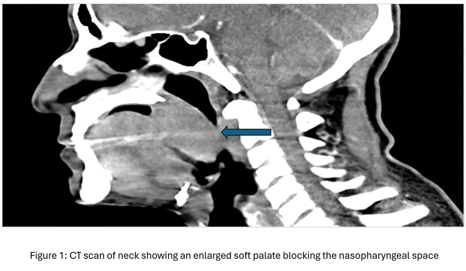
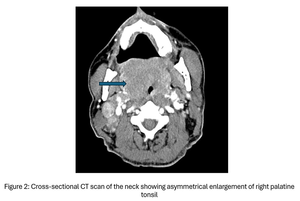
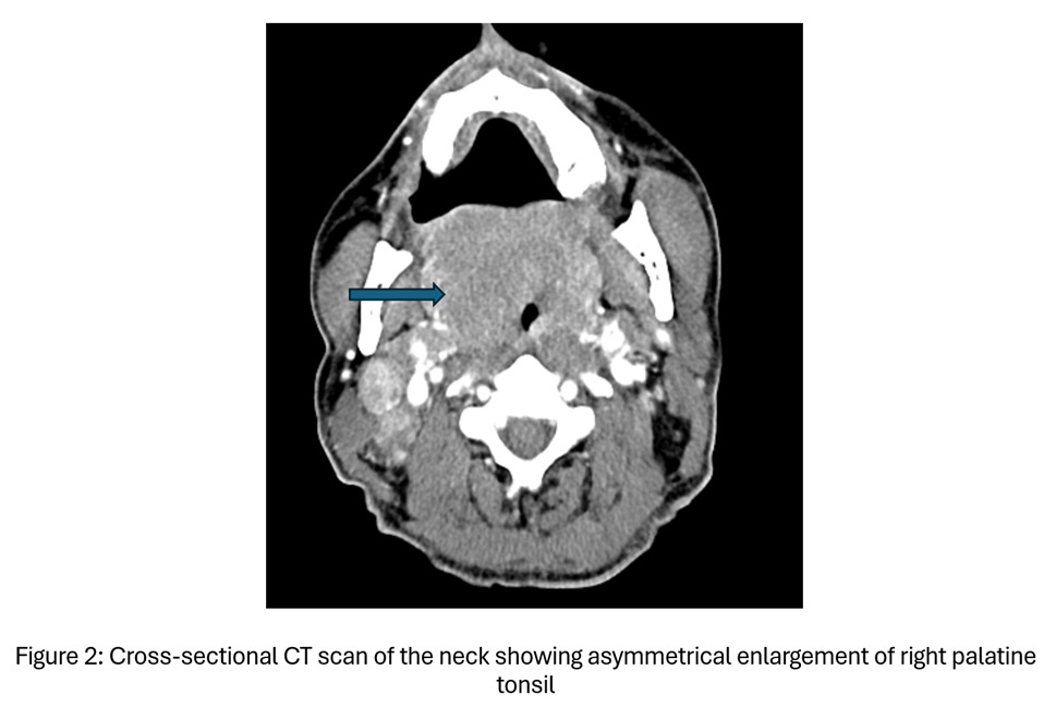
Initial laboratory evaluation revealed mild anemia (hemoglobin, 12.3 g/dL), hypokalemia (potassium, 3.0 mmol/L), proteinuria (70 mg/dL), mildly elevated inflammatory markers (erythrocyte sedimentation rate 23 mm/hr; CRP 1.77 mg/dL), normal hepatic and renal functions with a normal serum lactate level (1.9 mmol/L). Infectious evaluation, including a respiratory viral panel and Group A Streptococcus PCR, and HIV ag/ab were negative. Even though IgG antibody level for EBV was elevated, IgM antibody was negative. Additional evaluation for autoimmune and complement-mediated etiologies, including C3, C4, C1 esterase inhibitor, rheumatoid factor, and HLA-B27, were unrevealing. Contrast-enhanced computed tomography (CT) of the neck revealed asymmetric enlargement of the right palatine tonsil with a possible peritonsillar abscess and significant edema of the soft palate. The patient was treated empirically with ampicillin-sulbactam, dexamethasone, and famotidine. His symptoms improved, oral intake was restored, and he was discharged on hospital day 5 in stable condition.
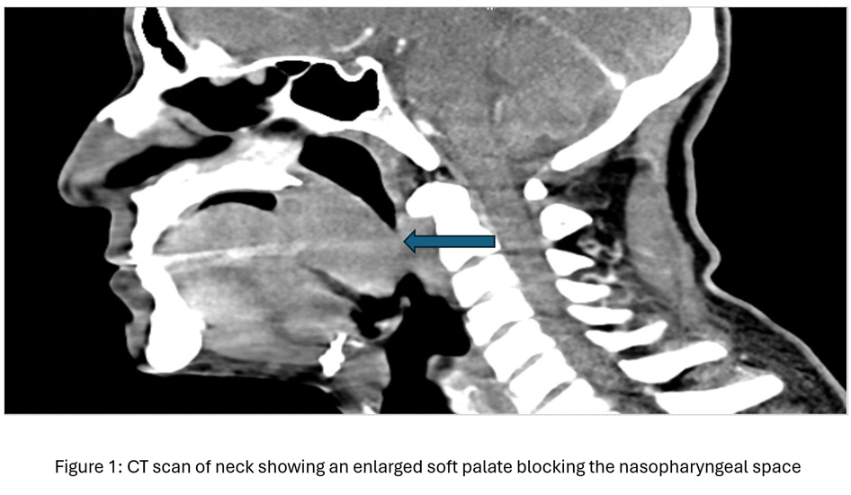
The patient returned 48 hours after discharge with worsening dysphagia and throat pain. Examination revealed marked enlargement of the soft palate and tonsils, raising concern for impending airway compromise, and nasotracheal intubation was performed. Initial laboratory evaluation revealed hyponatremia (127 mmol/L), hypokalemia (3.0 mmol/L), elevated lactate (6.3 mmol/L), mild transaminitis (AST 73 U/L), and thrombocytopenia (148 × 103/μL). Repeat contrast-enhanced CT of the neck demonstrated diffuse oropharyngeal mucosal thickening and hyperenhancement, bilateral level II lymphadenopathy up to 2.6 cm, and an absent right parotid gland of uncertain significance.
Because of hypotension and elevated lactate, possible sepsis was initially considered, and the patient was treated empirically with broad-spectrum intravenous antibiotics, including cefepime and vancomycin, as well as dexamethasone. His clinical status initially improved, allowing extubation after five days of mechanical ventilation. Because the etiology of the persistent pharyngeal edema remained unclear, a soft palate biopsy was obtained.
On hospital day six, the patient’s clinical course was complicated by worsening lactate levels, which were 6.3 mmol/L on admission and rose progressively to 7.0 and then 12.7 mmol/L despite aggressive fluid resuscitation and broad-spectrum antibiotics. At that stage, he did not require vasopressors; blood cultures were negative, and no clear source of infection was identified. These findings strongly supported a malignancy-associated type B component to the lactic acidosis; however, a mixed type A/type B process could not be excluded as hemodynamic instability subsequently developed. Concurrently, hepatic function deteriorated, with transaminases rising from baseline (ALT 33 → 320 U/L, AST 36 → 390 U/L), alkaline phosphatase increasing to 381 U/L, and LDH reaching 1,260 U/L. Abdominal ultrasonography revealed hepatomegaly with fatty infiltration and mild splenomegaly (13.2 cm). CT of the abdomen and pelvis with intravenous contrast showed an incidental 13 mm indeterminate pancreatic head cystic lesion.
On hospital day seven, the patient developed refractory hypoglycemia (glucose 24 mg/dL) requiring a dextrose 10% infusion, worsening lactic acidosis (anion gap 24, bicarbonate 6 mmol/L, and lactate level rising to 18.6 mmol/L), and worsening liver enzymes. He also developed visual hallucinations and progressive agitation, requiring re-intubation for airway protection and clinical decompensation. Within 12 hours, lactate rose dramatically from 18 to 45 mmol/L despite aggressive fluid resuscitation and bicarbonate infusion. Continuous renal replacement therapy was planned, and a trialysis catheter was placed; however, continuous renal replacement therapy could not be initiated because of severe, rapid hemodynamic instability.
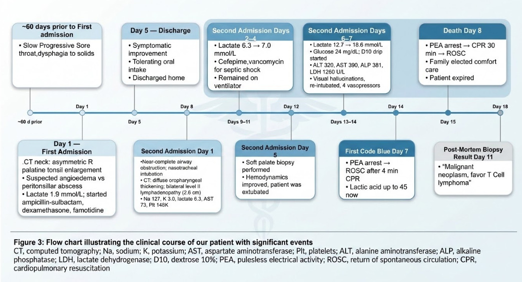
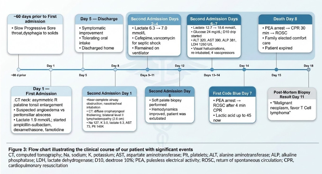
Despite maximal supportive care, including four vasopressors and continuous bicarbonate infusion, the patient suffered pulseless electrical activity (PEA) cardiac arrest on hospital day seven, with return of spontaneous circulation after four minutes of cardiopulmonary resuscitation (CPR). Within a few hours, a second PEA cardiac arrest occurred, and CPR was performed for approximately 30 minutes. Following discussion of goals of care, the family elected to forgo further resuscitative efforts, and the patient died shortly thereafter. Histopathologic examination and immunohistochemistry of the soft palate biopsy, reported three days after his death, demonstrated negative CD20 and diffuse positivity for CD3, favoring T-cell lymphoma. Because additional immunophenotyping, flow cytometry, molecular clonality studies, and staging could not be performed before the patient’s death, definitive WHO/ICC classification was not possible. Figure 3 shows the flow chart representing the patient’s clinical course.
Discussion
This case exemplifies several diagnostic challenges associated with oropharyngeal lymphoma, which frequently resembles benign infectious or inflammatory diseases.3 It parallels our previously published case of EBV-related lymphoma, wherein the diagnosis of aggressive B-cell lymphoma was delayed due to diagnostic anchoring to a benign viral illness.14 The delays in diagnosis can be attributed to three primary factors: the nonspecific symptoms that overlap with benign conditions, the empiric treatment that temporarily alleviates symptoms, particularly with steroids, and the low clinical suspicion of malignancy in the absence of classic red flags.4
In our patient, dexamethasone likely provided transient relief by reducing tumor-associated edema, reinforcing the impression of an inflammatory process. Corticosteroids can induce transient regression of lymphomatous tissue through cytolysis of lymphoma cells, potentially masking the underlying malignancy. Madireddy et al. [2025] reported that prebiopsy corticosteroid administration did not significantly reduce the diagnostic yield of DLBCL, with yields of 83% and 81%.15 However, these diagnostic-yield data are specific to DLBCL and cannot automatically be extrapolated to T-cell lymphoma. Clinical improvement following steroid therapy may still delay recognition and tissue diagnosis. Also, Gallogly et al. [2023] found that empiric antibiotic prescribing was associated with increased time to diagnosis of head and neck cancer. The patient received empiric broad-spectrum antimicrobial and corticosteroid therapy during both admissions, with only transient clinical improvement followed by recurrence and progression of the oropharyngeal process. As emphasized by Mills et al. (2023), recurrent or progressive inflammatory-appearing lesions despite empiric therapy should prompt clinicians to maintain a high level of suspicion for malignancy.7
Several clinical features in our patient, including asymmetric palatal enlargement, cervical lymphadenopathy, recurrent symptoms despite empiric therapy, and persistent oropharyngeal swelling, represented important clinical red flags. The patient exhibited asymmetric palatal enlargement, which a recent meta-analysis found to have 77% sensitivity and 96% specificity for malignancy, with the positive predictive value rising to approximately 38.5% when concurrent high-risk features, such as lymphadenopathy, are present.6 Bilateral cervical lymphadenopathy up to 2.6 cm was observed on repeat imaging; Fellner et al. [2020] found that lymphadenopathy was significant in the malignancy group among patients with unilateral tonsillar enlargement.16 Edwards et al. [2023] found that rapid unilateral tonsil enlargement was the only factor significantly associated with malignant outcomes in a series of 323 patients; this patient exhibited multiple such red flags.17
Acquired angioedema can occur in lymphoproliferative disorders, usually in association with C1-INH deficiency from increased consumption or anti-C1-INH autoantibodies.12,13 The available complement studies did not support classic C1-INH-deficient acquired angioedema; however, only quantitative C1-INH was assessed, and functional C1-INH deficiency therefore could not be excluded. Anti-C1-INH autoantibodies were not measured. Gunatilake et al. [2014] reported B-cell NHL with angioedema despite a normal C1-INH protein level, with reduced C1-INH function and low C1q and C4.18 Guidelines recommend assessment of both antigenic and functional C1-INH.19 Accordingly, true acquired angioedema could not be definitively confirmed or excluded in this patient; the angioedema-like appearance may instead have reflected lymphomatous infiltration of the soft palate.5
The clinical and metabolic findings strongly supported a malignancy-associated type B component to the lactic acidosis, consistent with the Warburg effect; however, a superimposed type A component could not be excluded after hemodynamic instability developed. The Warburg effect is aerobic glycolysis by malignant cells despite adequate oxygen, causing lactate overproduction. Liver infiltration with impaired hepatic clearance and cytokine-mediated dysfunction may cause hyperlactatemia. Rapidly progressive lactic acidosis, hypoglycemia, and hepatic dysfunction were consistent with malignancy-associated type B lactic acidosis. Van de Louw A et al. [2026] identified an association between lactic acidosis and mortality in ICU patients with hematologic malignancies, emphasizing etiology when lactate does not respond to resuscitation.20 Zhang W et al. [2025] reported that type B lactic acidosis due to lymphoma is misattributed to sepsis, with chemotherapy the sole independent predictor of survival (8). Chemotherapy was not initiated because tissue diagnosis was unavailable before deterioration.
Despite aggressive supportive measures, including intravenous fluids, bicarbonate infusion, vasopressor support, and planned renal replacement therapy, the patient’s metabolic derangements progressed relentlessly. This clinical course underscores that supportive therapy alone is insufficient to reverse malignancy-associated type B lactic acidosis in the absence of definitive treatment of the underlying malignancy. Wang et al. [2022] noted that alkali therapy has limited proven benefits in malignancy-induced lactic acidosis and may paradoxically exacerbate it by increasing net lactate production.8 In this case, each bicarbonate administration resulted in only transient hemodynamic improvement before blood pressure declined again, consistent with the literature indicating that without chemotherapy to halt tumor-driven glycolysis, supportive measures alone cannot reverse the underlying metabolic derangement8,9
Conclusion
The head and neck region is the second most common extra-nodal site for NHL. Oropharyngeal lymphoma should be considered in patients with persistent or recurrent asymmetric tonsillar or palatal enlargement, particularly when accompanied by cervical lymphadenopathy or failure to respond to empiric antimicrobial or corticosteroid treatments. Clinicians should also recognize rapidly progressive lactic acidosis without an identifiable cause of tissue hypoperfusion as a potential manifestation of malignancy-associated type B lactic acidosis (Warburg effect), as prompt diagnosis and initiation of chemotherapy remain the cornerstones of management and offer the greatest opportunity to improve outcomes.
Conflict of Interest
None of the co-authors has declared any conflict of interest.
Competing Interests
No competing interests were disclosed.
Funding
The authors did not receive support from any organization for the submitted work.
Consent to publish
Informed consent to publish has been obtained from the next of kin, as the patient is deceased.
Ethics
According to institutional policy, ethical review and approval are not required for this case report.
Author Contribution based on CRediT taxonomy
Conceptualization: Amrut Savadkar (Lead). Writing – original draft: Amrut Savadkar (Equal), Adeeba Sajid (Equal). Writing – review & editing: Amrut Savadkar (Equal), Ipsita Chauhan (Equal), Mansi Sharma (Equal), Suhasini Rallabandi (Equal), Pratibha Patil (Equal), Rahul Kashyap (Equal). Visualization: Amrut Savadkar (Equal), Adeeba Sajid (Equal). Supervision: Amrut Savadkar (Equal), Rahul Kashyap (Equal). Project administration: Amrut Savadkar (Lead).