1. INTRODUCTION
Primary myelofibrosis (PMF) is the rarest of BCR-ABL1-negative myeloproliferative neoplasms (MPNs), with an estimated incidence of 0.5 to 1.5 cases per 100,000 persons per year.1 It is characterized by clonal proliferation of hematopoietic stem cells, progressive bone marrow fibrosis, extramedullary hematopoiesis responsible for massive splenomegaly, and leukoerythroblastic anemia.
According to the 2022 WHO classification, the diagnosis of overt PMF requires: (i) megakaryocytic proliferation with reticulin/collagen fibrosis ≥ grade 2; (ii) a clonal marker — JAK2 V617F, CALR exon 9, or MPL W515 mutation, or, in their absence, a clonal cytogenetic abnormality recognized as an alternative criterion provided that other myeloid neoplasms have been excluded and the morphologic criteria are fulfilled; and (iii) at least one minor criterion: anemia, leukocytosis ≥ 11 × 10⁹/L, splenomegaly, or leukoerythroblastosis.2
Cytogenetic abnormalities are detected in 35–40% of PMF cases at diagnosis, the most common being del(20q), del(13q), trisomy 8, trisomy 9, and 1q duplication.3 Rearrangements involving 1q44 and 12q15 are exceptionally rare, and the translocation t(1;12)(q44;q15) has not, to our knowledge, been reported in any MPN in the indexed literature. We report herein a case of PMF at the osteomyelosclerotic stage harboring this novel clonal cytogenetic abnormality.
2. CASE REPORT
A 49-year-old man with insulin-dependent diabetes mellitus (HbA1c 11.1%) and untreated hypertension presented with a 4-month history of progressive fatigue and significant weight loss. Physical examination revealed massive splenomegaly reaching the umbilicus, confirmed by abdominal ultrasound (spleen dimensions: 22 × 10.5 cm, regular contours, homogeneous echotexture, with posterior displacement of the left kidney and a small intraperitoneal effusion). Liver size was normal with patent portal vein and hepatic veins. No deep lymphadenopathy was detected.
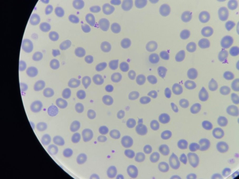
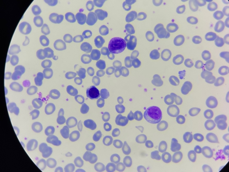
Complete blood count revealed: hemoglobin 8.0 g/dL, leukocytes 22,272/mm³, and platelets 698,000/mm³. Peripheral blood smear (MGG stain) showed numerous dacryocytes — a characteristic finding in myelofibrosis, although not specific, as teardrop cells may also occur in other marrow infiltrative disorders and in severe dyserythropoietic states — along with significant leukoerythroblastosis: 15% metamyelocytes and myelocytes, 15% circulating blasts, and 25 erythroblasts per 100 white blood cells (Figure 1, Figure 2). This presentation initially raised concern for acute leukemia or a blastic phase of chronic myeloid leukemia (CML).
._dacryocytes_with_marked_anisopoikilocytosis__charact.jpeg)
._myelemia_and_circulating_blasts_in_a_leukoerythroblas.jpeg)
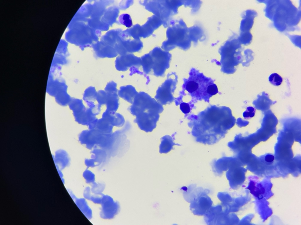
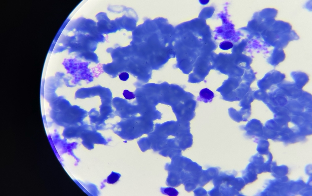
Bone marrow aspirate demonstrated hypocellular marrow (granulocyte/erythroid ratio 1.2), 6% blasts without overt dysplasia, atypical megakaryocytes with dense polylobulated nuclei in loose clusters, numerous activated macrophages, lymphocytes at 21%, and plasma cells at 2% (Figure 3, Figure 4). Bone marrow biopsy (1.4 cm trephine core) revealed demineralized bony trabeculae dissociated by dense collagen fibrosis, with overall cellularity approximately 10%, intrasinusoidal megakaryocyte clusters, and hypoplastic erythroid lineage. Gomori staining confirmed severe reticulin densification; Masson’s trichrome confirmed collagen fibrosis — both consistent with myelofibrosis grade 3 (MF-3). Immunohistochemistry for CD34 (clone QBEnd10) identified only 1% blasts, strongly arguing against overt blastic transformation. The combination of MF-3 fibrosis with a low in situ blast count favored a diagnosis of PMF at the osteomyelosclerotic stage, although the differential diagnosis with PMF with excess blasts (accelerated phase) was considered and is discussed below.
._dysplastic_megakaryocyte_with_dense_polylobulated_nucleu.jpeg)
._blasts_(6_)_within_a_lymphoplasmacytic_background.jpeg)
Multiparameter flow cytometry identified 13% myeloid blasts with the following immunophenotype: MPO-c+ (12%), CD34+ (92%), HLA-DR+ (87%), CD117+ (86%), CD13+ (66%), CD33+ (44%), CD38+ (46%), CD64+ (14%), with notable overexpression of CD123 (83%). B-lymphoid markers (CD19, CD22, CD10, CD20) and T-lymphoid markers (CD7, CD5, CD3, CD4, CD8) were all negative. Intracytoplasmic terminal deoxynucleotidyl transferase (TdT) expression was detected in 1% of cells. The discordance between the cytologic blast count on aspirate (6%), the flow cytometry blast fraction (13%), and the CD34 immunohistochemical blast fraction (1%) deserves careful interpretation. In severely fibrotic marrows, aspirate and flow cytometry samples are commonly hemodiluted by peripheral blood, which may inflate the apparent blast fraction; conversely, in situ CD34 immunohistochemistry on the trephine biopsy is the WHO-recommended reference method for blast enumeration in fibrotic marrows.2,4 Nevertheless, a 13% flow cytometric blast fraction is not negligible and must be weighed in the differential with PMF with excess blasts and with accelerated-/blast-phase PMF; in our patient, the low in situ CD34 blast count (1%) and the histopathologic pattern argued against overt blastic transformation but did not categorically exclude an early accelerated phase, a point that ideally would have warranted close longitudinal monitoring.
Conventional bone marrow cytogenetics (RHG banding, 15 metaphases analyzed, 24-hour culture) demonstrated the following karyotype:
46,XY,t(1;12)(q44;q15)[14]/46,XY
This pseudodiploid karyotype confirmed a balanced translocation between the long arm of chromosome 1 (q44) and the long arm of chromosome 12 (q15) in 14 of 15 metaphases, with one normal metaphase. Per PMF cytogenetic risk stratification, this was classified as intermediate risk.5 Importantly, an initial external cytogenetic interpretation had suggested compatibility with myelodysplastic syndrome or acute leukemia — a discordance resolved exclusively through systematic anatomocytogenetic confrontation with the bone marrow biopsy findings.
JAK2 V617F mutation testing was negative. CALR and MPL mutation testing, as well as next-generation sequencing (NGS), were not performed, which represents an important methodological limitation (further discussed in section 3.5). Under the 2022 WHO criteria, the documented clonal cytogenetic abnormality may serve as an alternative clonal marker once other myeloid neoplasms have been excluded and the morphologic criteria are fulfilled, as was the case here.2
IPSS prognostic scoring: constitutional symptoms (+1 point), hemoglobin < 10 g/dL (+1 point), circulating blasts ≥ 1% (+1 point) — total score 3, high-risk category, median expected survival approximately 27 months.6 Therapeutic evaluation for allogeneic hematopoietic stem cell transplantation and/or ruxolitinib was initiated.7 Unfortunately, the patient was subsequently lost to follow-up, and the planned therapeutic strategy could not be implemented; the clinical course beyond the initial diagnostic work-up could therefore not be documented.
3. DISCUSSION
3.1. The Diagnostic Pitfall
The clinicobiological presentation of this case — massive splenomegaly, hyperleukocytosis at 22,272/mm³, 15% circulating blasts, and hemoglobin 8.0 g/dL — strongly mimicked acute lymphoblastic leukemia or a blastic transformation of CML. The bone marrow biopsy, demonstrating MF-3 collagen fibrosis with a low in situ CD34 blast count (1%), made overt acute leukemia much less likely and was consistent with PMF at the osteomyelosclerotic stage.2,4 However, the differential diagnosis between PMF with excess blasts (10–19% blasts, accelerated phase) and overt blast-phase PMF (≥ 20% blasts) must be carefully considered in any patient with discordant blast counts between blood, aspirate, flow cytometry, and biopsy. In our patient, the convergence of MF-3 collagen fibrosis, characteristic megakaryocytic atypia, and a low in situ blast count favored overt PMF rather than accelerated- or blast-phase disease, while acknowledging that longitudinal monitoring would have been needed to formally rule out early accelerated-phase disease. This case illustrates that cytogenetic results should be interpreted in their histological context and that bone marrow biopsy is a key examination in this differential.
3.2. Clonality Established by Cytogenetics
With JAK2 V617F testing negative and CALR/MPL testing unavailable, clonality was supported by the t(1;12)(q44;q15) translocation present in 14 of 15 metaphases. Under the 2022 WHO classification, a clonal cytogenetic abnormality may serve as an alternative clonal marker once other myeloid neoplasms have been excluded and the morphologic criteria fulfilled.2 In our patient, the combination of MF-3 fibrosis, characteristic megakaryocytic atypia, low in situ blast count, and a clonal cytogenetic abnormality was compatible with the WHO criteria for overt PMF, while acknowledging that a complete molecular work-up (CALR, MPL, and broader NGS) was not feasible — a limitation that tempers the strength of the “triple-negative” interpretation.
3.3. Genomic Significance of the Implicated Regions
The t(1;12)(q44;q15) involves two poorly characterized genomic regions in MPNs. We emphasize that the following discussion is hypothesis-generating and is based on the genomic content of the cytogenetic bands rather than on direct molecular evidence of gene disruption or fusion in this patient. The 1q44 region harbors SMYD3, a methyltransferase implicated in oncogenesis through histone H3K4 methylation and epigenetic dysregulation, and AKT3, a serine/threonine kinase of the PI3K/AKT survival signaling pathway. The 12q15 region contains MDM2, a negative regulator of the tumor suppressor p53 with established roles in myeloid malignancies. The adjacent 12q14.3 locus harbors HMGA2, a myeloid oncogene whose overexpression has been reported in several MPNs, including PMF3,7; depending on the precise breakpoint location, a 12q14–q15 rearrangement could conceivably co-implicate HMGA2 through cis-regulatory disruption. The actual involvement of SMYD3, AKT3, MDM2, or HMGA2 in this patient remains entirely speculative in the absence of FISH or targeted NGS confirmation, which are warranted to characterize any putative fusion gene(s) or regulatory disruption.
3.4. Therapeutic Implications
Multiparameter flow cytometry showed marked CD123 expression on myeloid blasts. CD123 (IL-3 receptor alpha chain) overexpression is documented across several myeloid malignancies, but its current therapeutic actionability is established essentially in blastic plasmacytoid dendritic cell neoplasm, where tagraxofusp (SL-401) is approved,8 and in investigational settings for acute myeloid leukemia. CD123-directed therapy does not currently constitute a validated therapeutic indication in PMF or in PMF-related blastic transformation outside clinical trials. We therefore report this finding as a biological observation that may inform future investigational strategies rather than as a current clinical opportunity.
3.5. Limitations
Several limitations must be emphasized. First, CALR and MPL mutation testing and NGS were not performed; this significantly limits the strength of the “triple-negative” interpretation, which is currently supported only by a negative JAK2 V617F assay. Second, FISH and targeted NGS on the 1q44 and 12q14.3–q15 breakpoints have not been performed, so the putative fusion gene(s) and any actual gene disruption remain entirely hypothetical at the molecular level. Third, prognostic stratification relied on the original IPSS6; contemporary scoring systems such as DIPSS-plus and MIPSS70+ v2.0, which incorporate molecular data, could not be applied. Fourth, the patient was lost to follow-up after the initial diagnostic work-up; longitudinal clinical, hematologic, and cytogenetic data were therefore not available, which precludes any inference regarding disease course, response to therapy, or risk of blastic transformation.
4. CONCLUSION
This case report highlights several clinically and scientifically relevant lessons in the diagnosis and management of advanced PMF.
From a diagnostic standpoint, the initial presentation illustrates a well-recognized pitfall: osteomyelosclerotic PMF can convincingly masquerade as acute leukemia or blastic CML transformation. Bone marrow biopsy with MF-3 fibrosis grading and CD34 immunohistochemistry remains a key reference examination for addressing this differential diagnosis, and cytogenetic results should be interpreted in their histological context.
From a diagnostic-criteria standpoint, this case illustrates that, once other myeloid neoplasms have been excluded and the morphologic criteria are met, conventional karyotyping can contribute meaningfully to the diagnosis of triple-negative MPN by providing an alternative clonal marker under the 2022 WHO criteria. This supports the practice of performing conventional cytogenetics in suspected MPN whenever driver mutation testing is negative or incomplete.
From a scientific standpoint, the t(1;12)(q44;q15) with breakpoints at 1q44 and 12q15 has not, to our knowledge, been previously described in PMF or in any MPN in the indexed literature. The potential involvement of SMYD3, AKT3, MDM2 — and possibly HMGA2 in the case of an extended 12q14.3–q15 breakpoint — is purely hypothetical at this stage and would require FISH and targeted NGS to be substantiated.
Finally, the marked CD123 expression on myeloid blasts is a biological observation rather than a current therapeutic indication; CD123-directed therapies are presently approved only in BPDCN and remain investigational in acute myeloid leukemia, with no established role in PMF outside clinical trials. This finding nonetheless underscores the value of systematic immunophenotyping in advanced myeloid disorders.
Acknowledgments
The authors would like to express their sincere gratitude to all individuals and institutions who provided support throughout this research.
Authors’ Contribution
Conceptualization: Dadi Chaymae; Methodology: Dadi Chaymae; Formal Analysis: Dadi Chaymae; Investigation: Dadi Chaymae; Data Curation: Dadi Chaymae; Writing – original draft: Dadi Chaymae; Writing – review & editing: Dadi Chaymae, Sami Zarhloul, Youssef Bighouab, Yousra Manar; Validation: Abdelkader Belmekki; Supervision: Abdelkader Belmekki.
Funding
None
Patient consent
Written informed consent was obtained from the patient for publication of this case report and all accompanying clinical images.
Competing Interests
No competing interests were disclosed.
Informed Consent Statement
All authors and institutions have confirmed their approval for publication of this manuscript.
Data Availability Statement
All data are available upon reasonable request and limitations.
STATEMENT FOR STUDIES INVOLVING HUMAN SUBJECTS
This case report was conducted in accordance with the ethical principles set forth in the Declaration of Helsinki. Written informed consent was obtained from the patient prior to the collection of clinical data and images for publication purposes. Patient confidentiality was preserved throughout the manuscript. No interventional procedures beyond standard clinical care were performed for the purpose of this report. Formal institutional ethics committee approval was not required for a single observational case report at our institution.