Background
Blastic plasmacytoid dendritic cell (pDC) neoplasm (BPDCN) is a rare aggressive hematologic malignancy derived from pDCs. This malignancy is characterized by heterogenous clinical features, complex diagnostic process and numerous changes in nomenclature and classification over time. pDCs play a central role in antigen presentation and the regulation of adaptive immune responses.1–3 BPDCN is characterized by poor prognosis, with a median overall survival of 8–14 months and poses a significant diagnostic challenge due to overlapping morphologic and immunophenotypic features with other hematologic neoplasms, particularly mature pDC proliferations (MPDCP) associated with myeloid neoplasms.4,5 Increased recognition of BPDCN in recent years is largely attributed to its distinctive clinical presentation, most notably prominent cutaneous involvement. In these cases, the distinction between BPDCN and MPDCP is critical and often challenging. BPDCN is typically defined by co-expression of CD4, CD56, and CD123, along with markers such as TCL1, CD303, and CD304, and usually lacks myeloid lineage markers (e.g., MPO, CD13, CD33). A hallmark feature is prominent skin involvement with infiltration of immature blastic cells deeply into the dermis, often representing the initial disease manifestation.4,5 In contrast, MPDCP is characterized by the expansion of mature pDCs with a distinctive immunophenotypic profile. These cells typically express high levels of CD123, HLA-DR, CD303, and CD304, along with CD4 and TCL1, while lacking CD56 and lineage-specific markers of B cells, T cells, and myeloid blasts. In contrast to BPDCN, MPDCP usually demonstrates a more mature immunophenotype and is commonly associated with an underlying myeloid neoplasm such as myelodysplastic syndrome, chronic myelomonocytic leukemia (CMML), or acute myeloid leukemia.6
Case Presentation
A 76-year-old man with a history of prostate cancer and previously diagnosed CMML in 2013, treated with azacitidine at an external medical institute. No molecular or cytogenetic data from that time are available, as such analyses were not performed or documented. Accordingly, there is no information regarding myeloid-associated mutations from the initial CMML diagnosis. The patient was subsequently referred to our institute in September 2024 following the development of cutaneous lesions for further diagnostic evaluation. The lesions initially appeared on the scalp and subsequently spread to the trunk and extremities.
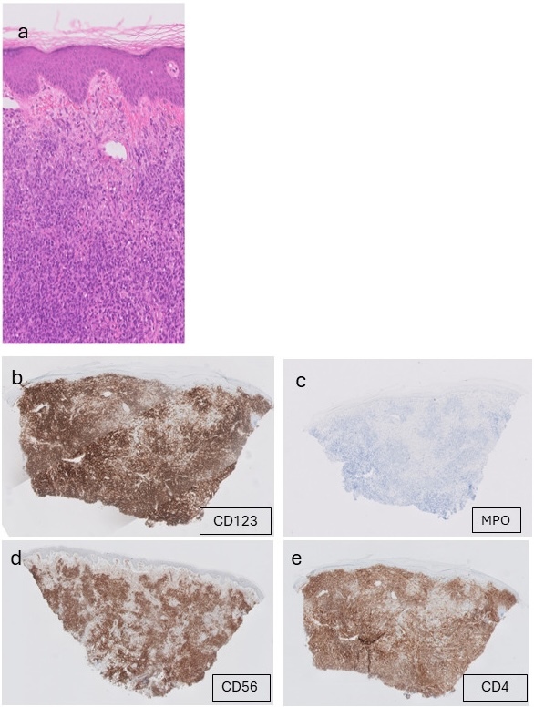
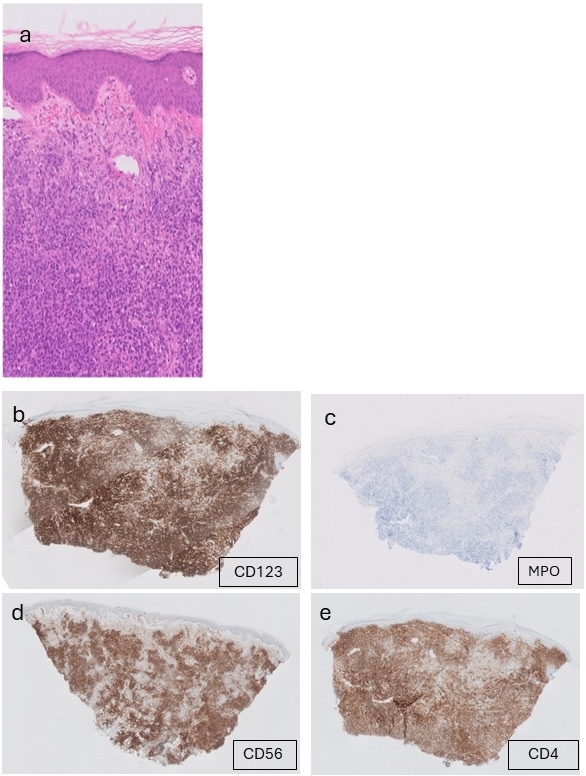
Dermatologic examination revealed multiple violaceous plaques and nodules with diffuse distribution, consistent with the classic cutaneous presentation of BPDCN (Figure 1). Skin biopsy demonstrated diffuse dermal infiltration by monomorphic blastoid cells (Figure 1a). Immunohistochemistry showed positivity for CD123 (b), CD56 (d), and CD4 (e), weak positivity for CD117 and TdT, and negativity for CD34 and MPO (c), supporting a diagnosis of BPDCN. A proliferation index of approximately 50% was observed by biomarker Ki-67.
In December 2024, the patient was referred for hematologic evaluation due to clinical deterioration, including severe anemia (hemoglobin 7 g/dL), thrombocytopenia, and renal dysfunction.
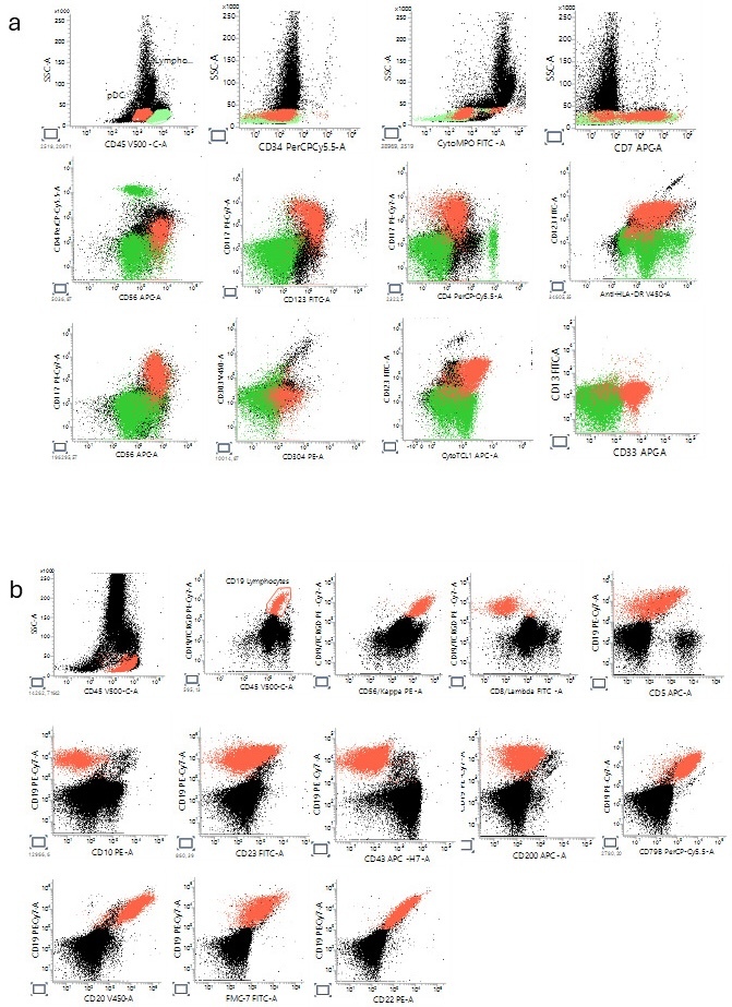
There was an increase in blast cells in suboptimal and non-diagnostic bone marrow aspiration due to hypocellularity. Normal hematopoiesis was not observed. By multiparameter flow cytometry a blast population expressing CD4 (dim), CD56, CD123, CD304 and cytoplasmic TCL1, with negative CD303 expression was identified (Figure 2a).
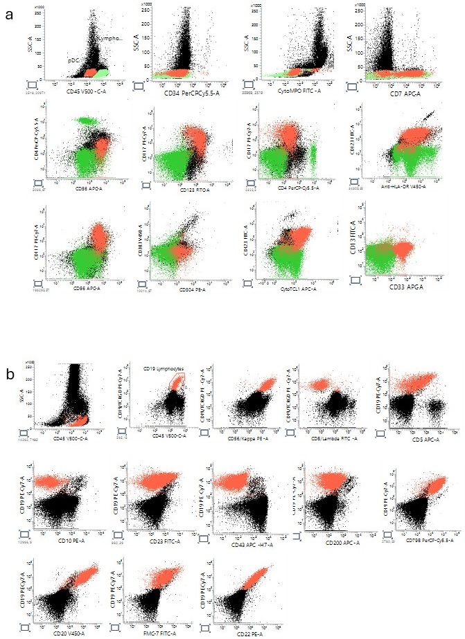
At this point, two main differential diagnoses were recognized. The differential diagnosis between BPDCN and MPDCP was challenging due to the partially overlapping immunophenotype. Flow cytometry confirmed expression of CD4, CD56, CD123, the classical triad in the BPDCN entity, along with the expression of CD304 and cytoplasmic TCL1, supporting pDC differentiation. However, these aberrant cells also expressed myeloid-associated markers CD117 and CD33, while lacking CD34, cytoplasmic MPO, and CD13. The population was negative for lineage-defining lymphoid markers (CD19, cytoplasmic CD79a, CD3, cytoplasmic CD3, CD10) and monocytic markers (CD14, CD64, CD15, CD11c), excluding acute lymphoblastic leukemia and monocytic differentiation. Considering the prominent cutaneous involvement, diagnosis strongly favored BPDCN rather than MPDCP, despite the absence of CD303 and the presence of CD117/CD33 expression.1,4–6 Cerebrospinal fluid analysis showed no evidence of central nervous system (CNS) involvement.
Cytogenetic and Genomic Findings
To further characterize the disease, optical genome mapping (OGM) was performed and revealed an interesting genomic profile, including:
-
Trisomy 12
-
t(14;19)(p13.13;q32.12) involving the immunoglobulin heavy chain (IGH) locus
Rearrangements involving IGH and chromosome 19 are classically associated with BCL3 activation, defining a rare but distinct subset of aggressive B-cell lymphoproliferative disorders.9–11 This cytogenetic signature is most described in atypical chronic lymphocytic leukemia and other non-Hodgkin lymphomas and is strongly associated with concurrent trisomy 12, atypical immunophenotype, and an aggressive clinical course.11,12 Importantly, IGH-associated rearrangements are not characteristic of BPDCN, which typically exhibits complex but non–IGH-driven genomic alterations.10,11 The presence of this aberration therefore supports the existence of a distinct clonal B-cell population, rather than representing part of the BPDCN disease biology.11,12
Treatment and Clinical Course
The patient was treated with tagraxofusp, a CD123-targeted therapy.13 Bone marrow evaluation after approximately 20 days of therapy showed a marked clinical and hematologic response. There was no evidence of BPDCN by morphology or flow cytometry. Nevertheless, 25% of the cells were lymphocytes. These cells were medium-sized with round nuclei with nucleoli, homogenous chromatin and agranular cytoplasm. Erythropoiesis and myelopoiesis maturation proceeded orderly to completion with myeloid lineage cells to nucleated erythroid precursors in the bone marrow.
Figure 2b illustrates flow cytometry immunophenotyping of the malignant B-cell population, expressing CD79b, CD22, FMC7, CD20 and restricted to the expression of Kappa light chain. These B cells were negative for CD10, CD23, CD200, CD43, and lambda light chain. Bone marrow biopsy was immunohistochemically stained and found to be positive for: CD20, CD79, CD5, and cyclin D1and negative for CD10, CD23, LEF1 and SOX11. Thereby confirming the diagnosis of mantle cell lymphoma (MCL). A concurrent clonal B-cell population with immunophenotyping pattern of MCL was confirmed by flow cytometric analysis representing an additional hematologic malignancy.14 Due to treatment-related toxicity, therapy was temporarily withheld.
Follow-up bone marrow evaluations in February and April 2025 showed no evidence of BPDCN recurrence.
Discussion
This case highlights several clinically important aspects, often taking part in the clinical diagnostic process. First, it demonstrates BPDCN with atypical immunophenotypic features, including expression of myeloid-associated markers (CD117 and CD33) and absence of CD303, creating a diagnostic challenge with MPDCP. The differential diagnosis between BPDCN and MPDCP can be challenging due to overlapping immunophenotypic features. Key distinguishing features between BPDCN and MPDCP include: BPDCN frequently expresses CD56, while MPDCP is CD56-negative; BPDCN shows blastic morphology while MPDCP has mature, non-blastoid morphology; and BPDCN commonly presents with characteristic skin lesions and may involve the CNS, whereas MPDCP rarely involves skin and does not involve the CNS.4,5 In the present case, the neoplastic cells expressed a typical pDC-associated profile, including CD4, CD56, CD123, CD304, and cytoplasmic TCL1. Notably, aberrant expression of myeloid-associated markers, including CD117 and CD33, was also observed, while MPO, CD34, TdT, CD13, CD64, CD10, and CD22 were negative. Such immunophenotypic overlap has been described in BPDCN and underscores that the presence of limited myeloid antigen expression does not exclude this diagnosis. Importantly, integration of the immunophenotype with the overall clinicopathologic context, including the absence of definitive myeloid differentiation markers and the strong pDC signature, supported classification within the BPDCN spectrum. BPDCN typically expresses strong CD123 (bright), TCL1+, CD4+, and often CD56+. Usually lacking strong myeloid markers, BPDCN can sometimes show aberrant myeloid antigen expression (e.g., CD33, CD117). The skin biopsy findings further support the diagnosis of BPDCN, in conjunction with the characteristic immunophenotypic profile and the overall clinicopathologic context. The distinction between BPDCN and MPDCP should be based on an integrated clinicopathologic, immunophenotypic, and molecular assessment rather than on isolated marker expression alone. Reliance on single antigenic findings may be misleading due to known immunophenotypic overlap between these entities, and accurate classification requires evaluation of the overall disease context.
This underscores the limitations of relying on single-lineage marker paradigms and emphasizes the need for an integrated diagnostic approach. Second, the case illustrates an atypical coexistence of BPDCN with a lymphoid malignancy, specifically an MCL–like clonal population. While BPDCN is known to co-occur with myeloid neoplasms such as CMML or myelodysplastic syndrome, its association with non-Hodgkin lymphoma is exceptionally rare.
Third, and most notably, the incorporation of OGM revealed a t(14;19) translocation with concurrent trisomy 12, a genomic profile strongly associated with BCL3-driven B-cell lymphoproliferative disorders. This finding provides critical biological insight, confirming that the lymphoid component represents a genetically distinct clone, rather than phenotypic variation within BPDCN.
The identification of a lineage-incongruent IGH-associated rearrangement is particularly informative in this diagnostically challenging case.
To our knowledge, the coexistence of BPDCN along with aberrant B-cell lymphocytes that have a t(14;19)-associated IGH rearrangement and trisomy 12 has not been previously reported. This case might expand the spectrum of genomic complexity associated with BPDCN.
Importantly, as tagraxofusp specifically targets CD123, accurate diagnosis is critical. Misclassification in cases with overlapping features may lead to inappropriate treatment selection, directly impacting both efficacy and toxicity. This highlights the essential role of precise immunophenotypic and genomic characterization in guiding therapy.
Conclusion
BPDCN is a rare hematologic malignancy with a distinctive clinical presentation, most notably involving the skin. Accurate diagnosis requires comprehensive integration of clinical, immunophenotypic, and genomic data.
This case underscores:
-
the diagnostic complexity of BPDCN, particularly in distinguishing it from pDC-acute myeloid leukemia
-
the importance of multiparameter flow cytometry
-
the added value of advanced genomic tools such as OGM
The identification of a concurrent IGH-associated chromosomal rearrangement with trisomy 12, along with cyclin D1 overexpression suggests the presence of an aberrant lymphoid clone and expands the recognized spectrum of composite hematologic malignancies involving BPDCN.
Therefore, early and precise diagnosis is essential for appropriate therapeutic decision-making and improved clinical outcomes.
Funding
The authors received no specific funding for this work.
Authors’ Contribution - CRediT
Conceptualization: Chen Glait Santar (Lead). Data curation: Chen Glait Santar (Equal), Lucille Hayman (Equal). Formal Analysis: Chen Glait Santar (Lead). Investigation: Chen Glait Santar (Lead). Methodology: Chen Glait Santar (Lead). Visualization: Chen Glait Santar (Lead). Writing – original draft: Chen Glait Santar (Equal), Lucille Hayman (Equal), Ben Zion Katz (Equal). Writing – review & editing: Chen Glait Santar (Equal), Lucille Hayman (Equal), Ben Zion Katz (Equal).
Competing of Interest statement
None of the authors have any conflicts of interest to disclose.
Informed Consent Statement
All authors and our institution have confirmed this manuscript suitable for publication.
Data Availability Statement
All are available upon reasonable request.