Background
Nasal-type extranodal natural killer (NK)/T-cell lymphoma (ENKTL) is a subtype of non-Hodgkin lymphoma (NHL), formerly referred to as lethal midline granuloma. It is a rare and aggressive disease associated with a poor prognosis.1 It typically involves the nasal cavity and paranasal sinus and is strongly associated with Epstein Barr virus (EBV) infection.2,3 This disease is commonly observed among East Asians and South Americans.4,5 NK/T-cell lymphoma usually manifests as localised necrosis, angioinvasion and angio-destruction.4 Due to the aggressive nature of the disease and its close anatomical proximity, there is a high risk of ophthalmic involvement.
Orbital apex syndrome is defined as a cluster of signs and symptoms due to multiple cranial nerve involvement in the orbital apex region, caused by a disease process.6 These causes may include inflammatory, infection, trauma, tumour, iatrogenic and vascular conditions.6 The cranial nerves which are typically affected are the optic nerve (cranial nerve [CN] II), oculomotor nerve (CN III), trochlear nerve (CN IV), abducens nerve (CN VI), and the ophthalmic division of the trigeminal nerve (CN V1).7
In this report, we present a case of left orbital apex syndrome (OAS) secondary to pansinusitis, with an underlying undiagnosed ENKTL.
Case Report
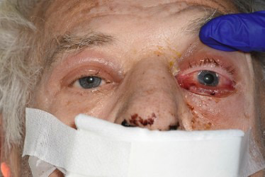
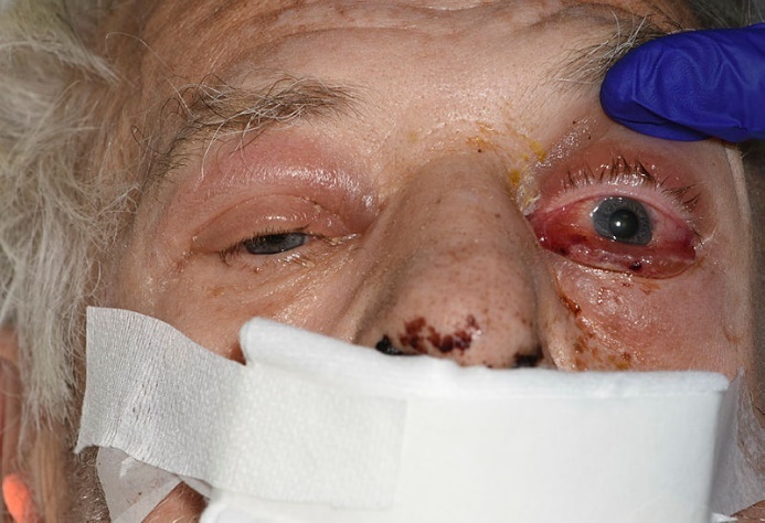
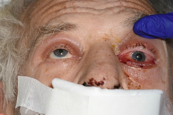
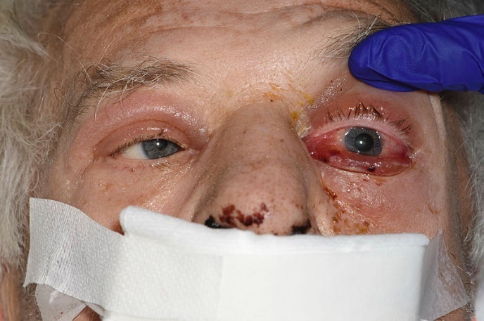
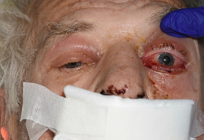
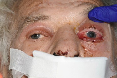
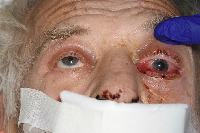
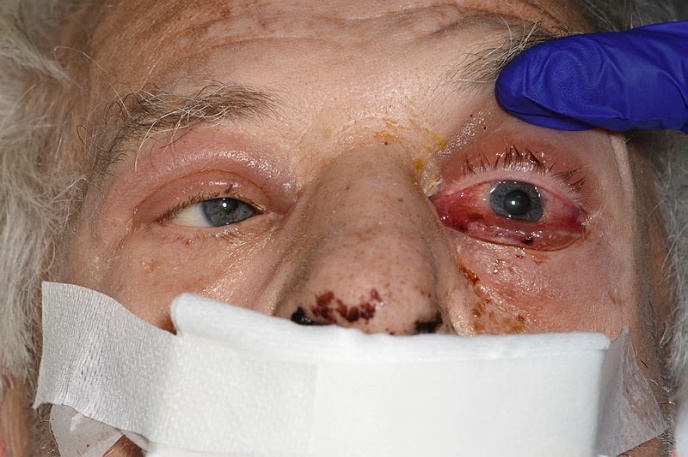
A 72-year-old Caucasian, otherwise healthy gentleman presented to the emergency department of a secondary care centre with a 6-month history of left nasal obstruction and 6-day history of left infraorbital nerve paraesthesia, reduced vision and restricted movement of the left eye. Initial ophthalmological examination revealed left eye complete ptosis, proptosis with firm retropulsion, ophthalmoplegia, fixed dilated pupil, relative afferent pupillary defect (RAPD), as well as efferent pupillary defect (Figure 1).
Computed tomography (CT) of the head demonstrated severe sinus disease with loss of the cribriform plate and soft tissue material extending from the sinuses to the inferior surface of the frontal lobes. Based on the findings from the magnetic resonance imaging (MRI) of the head, he was transferred to a tertiary centre with suspicion of possible mucormycosis. The MRI findings were reported as most consistent with an inflammatory aetiology. In particular, mucormycosis was suggested as a differential diagnosis given the presence of non-enhancing (necrotic) mucosa with extension into the retro-antral fat.
His inflammatory markers were mildly raised on admission (C-reactive protein 64mg/dL, white cell count 5.7x10⁹/L, neutrophils 3.76x10⁹/L) and LDH level was raised at 930 U/L. Other immunosuppression screens were negative, including HIV, hepatitis C antibody, hepatitis B surface antibody, hepatitis B surface antigen (HBsAg) and vasculitis screen (anti-protease antibody, PR3 and anti-myeloperoxidase antibody, MPO). Following initial discussion with the microbiology team upon admission to the tertiary centre, he was started on intravenous meropenem and liposomal amphotericin B.
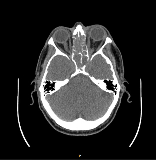
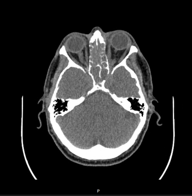
Urgent CT of the sinus with contrast showed pansinusitis, leading to blockage of the osteo-meatal complex, frontal recess and sphenoethmoidal recess bilaterally. Erosion was observed in the cribriform plate, nasal septum, as well as the middle and superior turbinates, and uncinate process on both sides, as well as the left sphenoid sinus and orbital apex. Figure 2 shows the lesion involving the left orbital apex, with associated erosion of the adjacent sphenoid sinus and orbital apex. A CT venogram showed no evidence of cavernous sinus thrombosis. However, initial MRI of the head revealed differential enhancement with associated bulging of the left cavernous sinus, findings that were highly suspicious for thrombosis. Following multidisciplinary discussion with the neurology and neuroradiology teams, who reviewed the imaging, it was concluded that thrombosis of the left cavernous sinus seemed likely, secondary to the underlying infection. Given the unilateral nature of the thrombosis, initiation of low-molecular-weight heparin was recommended. The initial working diagnosis was left OAS secondary to pansinusitis.
He underwent an emergency bilateral functional endoscopic sinus surgery, frontal balloon dilatation and left orbital apex decompression. Biopsies of abnormal granulation tissue were taken from the turbinates, sphenoid and nasal septum. Pathology results revealed markedly ulcerated nasal mucosa with marked infiltration by atypical lymphoid cells. The atypical lymphoid cells were extensively CD3+ T cells with CD30+ and Bcl-2+ staining. Ki-67 staining indicated a high proliferative index. The appearances favoured a high-grade T-cell NHL.
Further review by an external pathologist reported multiple tissue fragments, some of which were partially necrotic and infiltrated by atypical lymphoid cells. These cells were intermediate in size, with irregular nuclei, relatively abundant cytoplasm, and had high proliferative rate. The immunophenotype was consistent with an NK/T-cell lineage, with focal expression of EBV latent membrane protein 1 (LMP1). Immunohistochemistry for key ENKTL markers demonstrated CD2 positivity, variable CD3 expression, and CD56 positivity. Fluorescence in situ hybridisation revealed EBV in situ hybridisation positivity, with the majority of lymphoid cells positive for EBV using the Dako Epstein–Barr virus (EBER) peptide nucleic acid probe. Clonal T-cell receptor rearrangements were identified within a background of polyclonal T cells. Molecular analysis further demonstrated T-cell clonality, EBV positivity, and the presence of STAT3 and TP53 mutations.
Culture results from the nasal swab taken on admission were positive for Staphylococcus aureus and S. lugdunensis (a coagulase-negative Staphylococcus species that behaves similarly to methicillin-sensitive S. aureus). No fungal growth was detected after five days of incubation. Consequently, intravenous meropenem and liposomal amphotericin B were discontinued, and antimicrobial therapy was de-escalated to high-dose intravenous flucloxacillin.
A diagnosis of ENKTL nasal type, with EBV positivity and STAT3 and TP53 mutations, was made. A positron emission tomography-computed tomography (PET-CT) scan was performed for staging and demonstrated a very highly FDG-avid lesion centred in the nasal cavity, extending to the left maxillary sinus, ethmoid air cells, sphenoid sinus, and frontal sinus, with an SUVmax of 26.5. The scan also revealed extensive metabolically active disease with both nodal and extranodal involvement. Compared with the initial CT and MRI of the head obtained at admission, the findings indicated morphological progression over a three-week period. There was widespread disease affecting multiple distant organs, including the lungs, liver, spleen, adrenal glands, and the skeleton.
Unfortunately, the patient’s condition continued to deteriorate clinically. He subsequently developed a seizure a few days prior to the planned initiation of treatment. A CT scan of the head revealed spontaneous intracranial haemorrhage. Following multidisciplinary discussions between the medical, haematology, and intensive care unit teams, it was agreed that a palliative approach would be in the patient’s best interests. He succumbed to his illness before the initiation of chemotherapy. The planned treatment regimen consisted of gemcitabine, L-asparaginase, and oxaliplatin (GELOX).
Discussion
NK-cell malignancy is categorized by two types according to the World Health Organization: (i) ENKTL, i.e., nasal type, and (ii) aggressive NK-cell leukaemia.8 ENKTL is a rare and aggressive malignancy that primarily affects the nose and upper aerodigestive tract.9 Less commonly, it manifests as extranasal disease (with a poor prognosis), involving sites such as the skin, gastrointestinal tract, and salivary glands.10,11 ENKTL is predominantly observed among Asian and South American populations and is consistently associated with EBV infection in tumor cells.12 EBV is a key driver of lymphomagenesis in nasal-type ENKTL, with latent viral infection promoting malignant transformation through dysregulation of cell survival, proliferation, and the host immune response.13
Due to the close anatomical proximity, ocular complications are likely with nasal-type ENKTL. Hon et al.14 investigated the incidence and pattern of ophthalmic involvement in nasal NK/T-cell lymphoma, finding that six of 24 patients experienced vision-threatening complications, including uveitis/vitritis and orbital infiltration. Lance et al.15 reported a comparable case in a 78-year-old Vietnamese woman who presented with a chronic history of left-sided ophthalmic symptoms without signs or symptoms of sinusitis. Her CT of the head revealed extensive opacification of the left maxillary and ethmoid sinuses.15 Another case study by Cimino et al.2 described a patient diagnosed with stage IVA ENKTL nasal type, following chronic maxillary sinusitis. She later developed anterior uveitis and underwent treatment, but unfortunately, she died a year later. These case histories are summarised in Table 1.
Orbital apex syndrome (OAS), also known as Jacod syndrome, is characterized by impaired vision resulting from optic neuropathy combined with its predominant feature - total ophthalmoplegia.16 OAS involves the posterior-most portion of the orbit, the orbital apex, including the optic canal and the superior orbital fissure.17 Other important clinical features include proptosis, ptosis, and numbness or pain over the forehead.16 OAS has a broad differential diagnosis, including infectious, vascular, and neoplastic causes. Within the latter causes, lymphomas are an important consideration, as a range of NHL subtypes can involve the orbital apex and present with similar clinical features. This is illustrated by Borchard and Nayak,18 who reported a case of OAS secondary to diffuse large B-cell lymphoma, in which rapidly progressive ophthalmoplegia and visual loss underscored the aggressive nature of lymphomatous involvement.
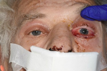
Our patient presented with a background of acute-on-chronic sinusitis and an acute onset of left-sided ocular symptoms. These included complete left eye ptosis, proptosis with firm retropulsion, and ophthalmoplegia involving the oculomotor nerve, trochlear nerve, and abducens nerve. He also had a fixed dilated pupil with an efferent pupillary defect (due to involvement of the parasympathetic fibres of the oculomotor nerve), as well as an RAPD consistent with optic nerve involvement. These findings are illustrated in Figures 3–6.
In addition, he also reported altered sensation in the left infraorbital nerve region, which could be explained by cavernous sinus syndrome (CSS), a condition with a similar presentation to OAS, with classical involvement of maxillary division of the trigeminal nerve (CN V2) and the oculosympathetic fibres.19 CSS affects the cavernous sinus, which lies posterior to the orbital apex.20 The cranial nerves typically involved include CN III, IV, VI, and the ophthalmic division of the trigeminal nerve (CN V1), with frequent involvement of CN V2 and sympathetic fibres.20 Unlike OAS, CSS does not usually involve the optic nerve unless the disease extends to the orbital apex.16 CSS mainly occurs secondary to cavernous sinus thrombosis, inflammation, infection, tumour and trauma.
The prognosis of ENKTL is generally poor, mainly due to its rapid progression and aggressive nature. Additionally, its rarity limits the availability of research and clinical studies. However, an increasing number of studies and trials, particularly in Asian countries, are being conducted to develop precise prognostic models and effective treatment strategies. One of the earliest models for predicting survival rates in patients with aggressive NHL is the International Prognostic Index.21 Other prognostic models include the Korean Prognostic Index, Prognostic index for NK/T-cell lymphoma (PINK) and PINK-EBV (PINK-E).22,23 Key factors associated with poor prognosis include age over 60 years, advanced disease (stage III/IV), non-nasal involvement on presentation, positive distant lymph node, and quantifiable EBV DNA at diagnosis.23 Presence of EBV DNA post-treatment is a significant prognostic factor for poor outcome.22
Based on the clinical and imaging findings of our patient, he had a PINK score of 3, reflecting age >60 years, stage IV disease, and distant nodal involvement, placing him in the high-risk prognostic group. Although tumour tissue demonstrated EBER positivity, plasma EBV DNA levels were not available; therefore, a formal PINK-E score could not be fully calculated. A key learning point from this case is that suspected mucormycosis should be evaluated within the broader clinical context, with emphasis on recognised immunosuppressive risk factors, rather than relying solely on radiological findings.
Asparaginase-based regimens remain the cornerstone of treatment for ENKTL, as asparaginase induces apoptosis in NK/T-cells through asparagine depletion.24 The 2025 European Society for Medical Oncology – European Haematology Association (ESMO-EHA) Clinical Practice Guideline recommends anthracycline-free, L-asparaginase-containing regimens as the standard of care for ENKTL.25 Combined radiotherapy and chemotherapy are preferred for limited-stage disease, whereas advanced-stage disease is treated with multi-agent systemic therapy such as DDGP (dexamethasone, cisplatin, gemcitabine, and pegaspargase), modified SMILE (dexamethasone, methotrexate, ifosfamide, pegaspargase, and etoposide), or P-GEMOX (pegaspargase, gemcitabine, and oxaliplatin).25 The guideline also recognises the emerging role of PD-1 inhibitors, particularly sintilimab in combination with P-GEMOX, as a promising first-line option for advanced ENKTL, although further validation and regulatory approval are awaited.25 This is supported by a recent single-arm study conducted by Tian et al.,26 who reported that the combination of sintilimab with P-GEMOX may represent an active and safe first-line option for advanced ENKTL. Table 2 summarises the ESMO-EHA guideline recommendations for ENKTL.
Combining modern radiotherapy and a non-anthracycline regimen containing L-asparaginase-based chemotherapy seems to achieve remission in advanced stage or relapsed disease.27 This evidence is supported by an extensive review conducted by Reneau et al.28 who analysed multiple studies on later-stage treatment. Although the SMILE regimen has demonstrated high efficacy, it is associated with significant toxicity.28 Consequently, less intensive regimens such as GELOX are often favoured in clinical practice due to their more favourable tolerability while maintaining good clinical efficacy, though it is usually for patients with stage I/II ENKTL.29 In this case, the GELOX regimen was selected over SMILE in view of the patient’s clinical condition, although SMILE may be considered in selected patients with advanced ENKTL who are able to tolerate more intensive therapy.
Overall, ENKTL with orbital apex involvement poses significant diagnostic and therapeutic challenges, highlights the importance of early recognition and the need for continued refinement of multimodal treatment strategies to improve clinical outcomes.
Conclusion
This case reinforces that suspected mucormycosis should be assessed within the broader clinical context, with emphasis on recognised immunosuppressive risk factors rather than radiological features in isolation. ENKTL, nasal type, is an aggressive malignancy that may initially mimic severe sinusitis. The presence of necrotic sinonasal disease, bony destruction, and rapidly progressive orbital or cranial neuropathies should prompt early biopsy to exclude underlying malignancy. Diagnosis requires histopathological evaluation with immunohistochemistry and EBV confirmation within tumour cells using EBER in situ hybridisation, as EBV serology alone is insufficient. Although rapid multidisciplinary management is essential, prognosis remains poor in advanced-stage disease, highlighting the importance of early recognition and timely diagnostic evaluation. Emerging therapies, including immunotherapies and novel combination regimens, show promise but further research and clinical studies are needed to improve outcomes in this aggressive disease.
Acknowledgment
The authors would like to thank the staff of the Ear, Nose and Throat and Ophthalmology Departments at the Lancashire Teaching Hospitals for their assistance in the clinical management of the patient. We sincerely thank the patient for providing consent to share his clinical details and images for educational purposes.
Ethical approval
Written informed consent was obtained from the patient for publication of any potential identifiable images or data included in this report.
Source of funding
The authors received no financial support for the report, authorship, or publication of this article.
Conflict of interest
The authors declare no conflict of interest.
CRediT Authorship Contribution Statement
Conceptualization: Farah Jeffry (Lead). Investigation: Farah Jeffry (Equal), Ding Yang (Equal). Project administration: Farah Jeffry (Lead). Visualization: Farah Jeffry (Equal), Ding Yang (Equal). Resources: Farah Jeffry (Lead). Writing – original draft: Farah Jeffry (Lead). Writing – review & editing: Farah Jeffry (Equal), Ding Yang (Equal). Formal Analysis: Ding Yang (Lead). Data curation: Gabrielle Thompson (Lead). Methodology: Gabrielle Thompson (Lead). Supervision: Rajeev Advani (Equal), John de Carpentier (Equal). Validation: Rajeev Advani (Equal), John de Carpentier (Equal).
Data access
Data and materials are available upon request.