Introduction
Chronic lymphocytic leukemia (CLL) is the most common form of adult leukemia worldwide, with approximately 22,000 new cases diagnosed annually, and accounting for roughly 4,400 deaths per year in the United States. The median age at diagnosis is 70 years, and the disease is defined by the presence of at least 5 × 10⁹/L monoclonal B lymphocytes in the peripheral blood.1 These malignant lymphocytes accumulate through impaired apoptosis rather than rapid proliferation, resulting in an expanding population of immunologically dysfunctional cells.2 The malignant clone typically co-expresses B-cell surface antigens CD5, CD19, and CD23, and often CD45 and CD200.2 Approximately 70% of patients are asymptomatic at diagnosis, with the disease first detected incidentally on a complete blood count revealing lymphocyte-predominant leukocytosis.1
Among the roughly one-third of patients who present with symptoms, 50% have generalized lymphadenopathy, 20–30% have hepatosplenomegaly, and up to 10% manifest constitutional B symptoms, hemolytic anemia, or hypogammaglobulinemia.1 Symptomatic patients may report abdominal discomfort, fevers, night sweats, unintentional weight loss, or fatigue.2
The past decade has brought remarkable advances in CLL treatment, including BCL-2 inhibitors, Bruton tyrosine kinase inhibitors, and anti-CD20 monoclonal antibodies.2 These therapies, however, further blunt both T-cell and B-cell humoral defenses, predisposing patients to infections from atypical bacteria, fungi, and herpesviruses.3 In early-stage and untreated disease, encapsulated bacteria are the predominant infectious pathogens, whereas in advanced or treated disease, opportunistic infections, including fungi and viruses, become increasingly prevalent.3
What is often underappreciated, however, is that CLL itself induces a state of immunodeficiency independent of any treatment. Innate immunity is compromised through impaired complement pathways and phagocytic dysfunction.3 CLL B cells promote immunosuppression via altered IL-10–mediated inhibition of immune response activation, a mechanism intrinsic to the disease rather than a consequence of chemotherapy or immunotherapy.4,5 Antiviral defense is further weakened by decreased plasmacytoid dendritic cell numbers and impaired IFN-α production, while T-cell exhaustion and defective activation and proliferation contribute to a profound state of immune dysregulation.5,6 As a result, patients may harbor significant immunodeficiency and remain vulnerable to opportunistic infections even in the absence of classic B symptoms or prior treatment. We present a case of opportunistic infection in a treatment-naïve CLL patient, underscoring the importance of recognizing CLL itself as an independent risk factor for immunocompromise in the hospital setting.
Case Report
An 89-year-old male with a past medical history of type 2 diabetes mellitus, aortic stenosis, osteoarthritis, and chronic kidney disease presented to the emergency department with one week of progressive lethargy. The patient had no prior history of malignancy, human immunodeficiency virus (HIV) infection, solid organ transplantation, chronic glucocorticoid use, or other known immunosuppressive conditions. Per collateral history obtained from the patient’s family, he had been in his usual state of health prior to this presentation and living independently.
On arrival, the patient was obtunded and unable to provide a history. Vital signs were notable for a temperature of 101.4°F (38.6°C), heart rate of 135 beats per minute, and blood pressure of 101/64 mmHg. Initial laboratory evaluation revealed a white blood cell count of 9.14 K/µL with an absolute lymphocyte count of 7.04 K/µL, hemoglobin of 7.7 g/dL with a mean corpuscular volume of 102.8 fL, markedly elevated creatine kinase of 4,517 U/L, creatinine of 2.1 mg/dL, and lactate of 2.5 mmol/L (Table 1).
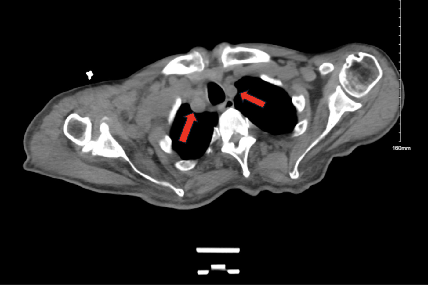
Urinalysis obtained on admission was unremarkable. Peripheral blood smear demonstrated small to intermediate lymphocytes with rounded nuclear contours, scant clear cytoplasm, irregular chromatin clumping, and scattered smudge cells. Computed tomography of the chest, abdomen, and pelvis revealed bilateral supraclavicular, axillary, and mediastinal lymphadenopathy, the largest measuring up to 2.7 cm in the right supraclavicular fossa suggesting of an underlying lymphoproliferative neoplasm (Figure 1).
Intravenous fluid resuscitation was initiated, and empiric broad-spectrum antibiotics utilizing vancomycin and piperacillin-tazobactam were started for presumed sepsis of unclear etiology. On hospital day 2, given the unexplained encephalopathy and incidentally discovered lymphadenopathy, the clinical team pursued concurrent diagnostic evaluation with excisional lymph node biopsy and lumbar puncture. Cerebrospinal fluid (CSF) analysis demonstrated an opening pressure of 8 cmH₂O, white blood cell counts of 61/µL, protein of 105 mg/dL (elevated), and glucose of 87 mg/dL. Histopathological examination of the right supraclavicular lymph node biopsy with flow cytometry revealed a CD45-positive cell population comprising 79% of the total analyzed cells, with lymphocytes comprising 78.4% of the population. The T cells (CD3+) comprised approximately 12.5% of the total cells analyzed and showed no evidence of phenotypic abnormality. B cells (CD19+) comprised 65% of total cells analyzed and showed dim surface lambda light chain restriction with co-expression for CD45, CD19, CD20 (dim), CD5, and CD23 (moderate), while negative for CD10, CD38, and FMC-7. The surface kappa to lambda light chain ratio was 0.01:1. Together, this confirmed a new diagnosis of CLL. HIV-1 and HIV-2 serologies were negative, and serum immunoglobulin levels (IgG, IgA, IgM) were not deficient.
CSF multiplex polymerase chain reaction (PCR) panel returned positive for C. neoformans, HSV-1, and HHV-6, and on hospital day 3, Cryptococcal antigen testing became positive, representing a potentially triple opportunistic coinfection in a treatment-naïve patient with no previously recognized immunocompromising condition. Targeted antimicrobial therapy was promptly initiated with liposomal amphotericin B and flucytosine for Cryptococcal meningoencephalitis and intravenous acyclovir for HSV-1 and HHV-6 central nervous system (CNS) infections.
Despite aggressive intervention, the patient’s clinical trajectory continued to deteriorate, with progressive hemodynamic instability requiring escalation to vasopressor support. On hospital day 4, blood cultures returned positive for C. neoformans, confirming disseminated fungemia. Despite aggressive medical therapy, given the absence of clinical improvement, a goals-of-care discussion was held with the patient’s family. The decision was made to transition to a comfort-focused approach, and life-sustaining treatments were withdrawn. The patient died on hospital day 8.
Discussion
A comprehensive review of CLL diagnosis and management concluded that all CLL patients should be considered immunocompromised, irrespective of treatment status.1 Humoral immunity is impaired through both quantitative and qualitative defects: hypogammaglobulinemia results from reduced antibody production, while defective B-cell maturation and differentiation impair functional antibody responses. Even if hypogammaglobulinemia is not present, intrinsic B-cell function is compromised and renders the patient immunocompromised.1 Critically, this immunodeficiency is intrinsic to the disease itself and can manifest in treatment-naïve, early-stage patients; it is not solely a consequence of chemotherapy or immunotherapy.3–5
Once functionally immunodeficient, bacterial, viral and fungal opportunistic infections must be considered for any patient presenting signs of an infection. A literature review of cryptococcosis in malignancy published between 1970 and 2014 demonstrated that 82% of cases were associated with hematologic malignancies.7 It is a rare, but clinically important opportunistic infection in CLL.8 While the vast majority occur in patients receiving immunosuppressive therapy, three prior reports have documented cryptococcal infection in treatment-naïve CLL, suggesting that the underlying immune dysfunction of CLL alone is sufficient to permit invasive fungal disease.7,8
Diagnosing cryptococcosis in non-HIV patients presents a particular challenge. Compared to HIV-associated disease, non-HIV cryptococcosis often presents atypically, leading to lower clinical suspicion and diagnostic delay.9 This delay is clinically consequential: outcomes in non-HIV patients with cryptococcosis are significantly worse than in their HIV-positive counterparts.10 The definitive global guideline recommends that any patient with compatible symptoms be evaluated for cryptococcosis irrespective of immune status. The importance of maintaining a broad differential is further underscored by a multicenter study of 475 patients demonstrating that 90% of cryptococcal cases occurred in non-HIV patients, with cancer as the most common predisposing factor.9,10
HSV-1 reactivation in CLL has been well documented in patients treated with purine analogs, anti-CD52 monoclonal antibodies and Bruton tyrosine kinase inhibitors.11 A Swedish population-based analysis reported an HSV reactivation incidence of 0.8 per 1,000 CLL patients compared to 0.04 per 1,000 controls.11 In treatment-naïve patients, however, it is nearly unheard of. Five CLL patients had biopsy-proven HSV lymphadenitis; four received no antiviral therapy and none developed disseminated infection. Notably, only one had clinical cutaneous herpes at any time.12 Although CSF PCR remains the gold standard for the diagnosis of HSV encephalitis, its positive predictive value depends substantially on the pre-test probability of true HSV encephalitis. In our patient, the absence of supportive features such as temporal lobe–predominant imaging abnormalities, hemorrhagic CSF, or xanthochromia substantially lowered that pre-test probability.12 It is therefore possible that the HSV-1 PCR positivity in our patient represented subclinical viral reactivation or low-level viral shedding rather than a pathogenic contributor to his encephalopathy. However, given the patient’s rapid clinical deterioration and the inability to obtain confirmatory neuroimaging or electroencephalogram, active HSV-1 CNS disease could not be excluded, and empiric acyclovir therapy was appropriately initiated.
HHV-6 has a less well-characterized association with hematologic malignancies but is recognized as reactivating in immunocompromised hosts. This may have occurred in our patient as asymptomatic reactivation would be PCR- positive, but was unable to be confirmed without quantitative PCR or magnetic resonance imaging findings suggestive of limbic encephalitis. However, in patients with hematologic malignancies, HHV-6 reactivation can cause severe organ dysfunction, with particular tropism for the brainstem, hippocampus, and limbic system. The most severe complication is limbic encephalitis.13–15
Our patient represents a potential triple coinfection with C. neoformans, HSV-1, and HHV-6 in a treatment-naïve CLL host. He had no elevated lymphocyte counts on prior complete blood counts to point to early-stage unrecognized CLL diagnosis and despite negative HIV serology and non-neutropenic state, this patient developed rapid encephalitis, making it difficult to discern whether his symptoms were solely cryptococcal meningoencephalitis alone versus true polymicrobial CNS disease. It is worth noting that up to 15% of cryptococcal infections in immunocompromised patients often present with a polymicrobial infection.10 The progressive nature of his decline lends itself to a cryptococcal infection, though the patient’s CSF analysis included an opening pressure of 8 cmH₂O, atypical for cryptococcal meningoencephalitis where opening pressures of >25 cmH₂O are common. This was potentially attributable to the HIV-negative host as current literature supports atypical presentations in non-HIV cryptococcosis or perhaps caused by a concurrent viral encephalitis.8 On admission, the patient was presumed immunocompetent, with no apparent immunocompromising risk factors such as HIV, prior malignancy, glucocorticoid use, or solid organ transplant. While the clinical team’s aggressive evaluation of incidentally discovered lymphadenopathy allowed for intervention within 48 hours, it is important to recognize that in non-HIV patients, the time to diagnosis of opportunistic infections is typically significantly longer and is associated with worse outcomes.
This case carries several practical implications. Clinicians should maintain a high index of suspicion in any patient presenting with unexplained sepsis or encephalitis and incidentally discovered lymphadenopathy. Early tissue biopsy with concurrent infectious workup should be initiated, given that patients with CLL are at risk for opportunistic infections. For CLL patients who do present with CNS symptoms, even those who are treatment-naïve, early oncology and infectious disease consultation should take place for appropriate risk stratification and to expedite appropriate diagnostic evaluation. CSF analysis with multiplex PCR for bacterial, viral, and fungal pathogens, and cryptococcal antigen detection, along with bacterial and fungal cultures, should be considered. These actions could enable earlier identification of the causative organism, thereby tailoring treatment and optimizing patient outcomes.
Authors’ Contribution – per CRediT
M.N, M.M, and E.K. conceptualized the case report; M.R. and R.C wrote the draft manuscript; E.K. revised the manuscript. All authors approved the final manuscript.
Competition of Interest – COPE
No competing interests were disclosed.
Informed Consent Statement
All authors and institutions have confirmed this manuscript for publication. Written consent was received from the patient to publish this work.
Data Availability Statement
All data are available upon reasonable request.