Introduction
Autoimmune hemolytic anemia (AIHA) is a rare and heterogeneous disorder caused by autoantibody-mediated red cell destruction. Warm AIHA is the commonest subtype in adults, but a small minority of patients with clinically convincing immune hemolysis have a negative direct antiglobulin test (DAT), usually because of low-density, low-affinity, or non-IgG antibodies that are not detected by routine methods. This DAT-negative subgroup remains diagnostically challenging and often requires clinicopathologic correlation rather than reliance on serology alone.1–3
Standard treatment for warm AIHA begins with corticosteroids, with rituximab widely used in steroid-refractory or steroid-dependent disease. However, patients who fail steroids, rituximab, and conventional immunosuppression represent an unmet need. Daratumumab, an anti-CD38 monoclonal antibody, provides a biologically plausible rescue strategy because it targets CD38-positive plasma cells, including long-lived autoreactive plasma cells that may persist despite CD20-directed therapy, and may also exert broader immunomodulatory effects. This remains a therapeutic rationale rather than proof of causality in any single case.1,2,4,5
Case presentation
To preserve patient confidentiality, exact calendar dates are not reported in the case description. The first relevant diagnostic clinical event is designated as Day 1, and subsequent events are reported as Day or Month relative to this event.
A 69-year-old woman was under treatment for high-grade endometrioid adenocarcinoma of the endometrium. The diagnostic biopsy on Day 1 showed high-grade adenocarcinoma/endometrioid carcinoma favored. Surgical pathology after total abdominal hysterectomy, bilateral salpingo-oophorectomy, pelvic lymphadenectomy, and omentectomy on Day 6 showed FIGO grade 3 endometrioid carcinoma, pT1aN0, with 0/15 regional lymph nodes involved. Immunohistochemistry showed pMMR with intact MLH1, PMS2, MSH2, and MSH6 expression; ER and PgR positivity; and HER2 score 0.
She subsequently received concurrent cisplatin-based chemoradiotherapy during Months 2-4, followed by two doses of vaginal brachytherapy after radiotherapy. A PET scan performed during Month 7 reportedly showed complete metabolic response.
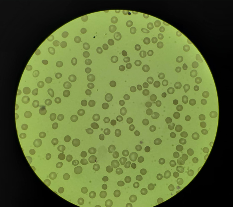
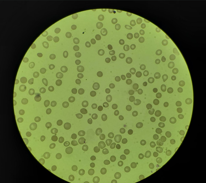
After oncologic treatment, she developed severe hemolytic anemia. The DAT was negative with an elevated reticulocyte count. PNH testing by flow cytometry was negative, and there were no schistocytes on peripheral smear. She was diagnosed with presumed DAT-negative autoimmune hemolytic anemia, with possible treatment-related or drug-related triggering. The course became steroid-dependent and ultimately refractory. Her comorbidities included systemic hypertension, type 2 diabetes mellitus, and hypothyroidism. There was no seizure history relevant to this case.
Hemolysis work-up
The available records supported ongoing hemolysis despite negative routine serology:
-
Direct antiglobulin test: negative
-
Indirect antiglobulin test: negative
-
Monospecific Coombs testing (IgG/IgM/C3d/C3c/IgA): negative
-
Haptoglobin: 0.027 g/L
-
Reticulocyte count: 5.6%; corrected reticulocyte count 2.7%
-
LDH: 307 U/L on Day 199 (local reference interval 140-280 U/L; previous available values: 319, 289, and 299 U/L)
-
Vitamin B12: 316 pg/mL
-
Stool occult blood: negative
-
Urine routine examination: within normal limits
-
Serum immunofixation: no monoclonal band
Bone marrow aspiration and biopsy performed on Day 132 showed erythroid prominence with micro-normoblastic to normoblastic erythropoiesis, few megaloblasts, and blasts approximately 0.1%; biopsy showed fragmented marrow with erythroid prominence, while plasma cells constituted approximately 7% of nucleated cells. Peripheral smear review during Month 7 described dimorphic anemia with polychromatophils and occasional nucleated red cells; neutrophil dysmorphology was interpreted as potentially treatment-related after prior chemo/radiation exposure rather than definitively clonal.
The diagnosis therefore remained presumptive rather than definitive. The constellation of low haptoglobin, reticulocytosis, mildly elevated LDH, marrow erythroid hyperplasia, absence of schistocytes, negative stool occult blood, and exclusion of paraproteinemia supported immune-mediated hemolysis, but overlapping therapy-related marrow injury or secondary marrow dysfunction could not be fully excluded.
Prior treatment
She received corticosteroids beginning on Day 116, followed by four doses of rituximab, with the fourth dose completed on Day 187. Because of ongoing disease activity, cyclosporine was started during Month 5, followed by mycophenolate mofetil from Day 244. Despite these sequential interventions, the anemia remained difficult to control.
Steroid dependence/refractoriness was operationally determined by the inability to maintain stable hemoglobin with corticosteroid-based therapy and by persistent or recurrent severe anemia despite subsequent rituximab and conventional immunosuppressive therapy. Exact steroid taper details were not fully available in the reviewed records.
Hemoglobin trend before daratumumab
The documented hemoglobin values showed marked fluctuation and recurrent severe anemia:
This pattern reflected persistent instability despite multiple lines of therapy.
Daratumumab treatment
Because of presumed multirefractory DAT-negative AIHA, subcutaneous daratumumab 1800 mg was introduced as salvage therapy. She received four doses in total; the fourth 1800-mg subcutaneous dose was administered on Day 288. The exact day numbers of the first three doses were not available in the reviewed chart.
At the Day 288 admission, she received daratumumab 1800 mg SC stat along with G-CSF 300 mcg SC stat because of falling total leukocyte counts. G-CSF was used for neutropenia/leukopenia support, and antimicrobial prophylaxis was prescribed because of neutropenia, cumulative immunosuppression, and expected infection risk with anti-CD38 therapy. The neutropenia was considered multifactorial, with possible contributions from recent chemoradiotherapy, marrow vulnerability, and immunosuppressive therapy; it was not attributed solely to AIHA.
No acute injection-related reaction was documented during the indexed administration. The chart available to us did not provide serial bilirubin, haptoglobin, LDH, or reticulocyte measurements after daratumumab. Pre-daratumumab transfusion exposure could not be completely reconstructed from the reviewed records; however, no red-cell transfusion requirement was documented after daratumumab in the available post-treatment follow-up.
Outcome and follow-up
A CBC on Day 304, after daratumumab exposure, showed:
-
Hemoglobin: 10.4 g/dL
-
RBC count: 3.13 million/uL
-
WBC count: 1.96 x 10^3/uL
-
Platelet count: 137 x 10^3/uL
-
Hematocrit: 31.9%
-
MCV: 101.9 fL
Thus, the most clinically important hematologic signal in the available follow-up was a rise in hemoglobin to 10.4 g/dL after failure of corticosteroids, rituximab, cyclosporine, and mycophenolate mofetil, with no post-daratumumab red-cell transfusion requirement documented. At the same time, macrocytosis, leukopenia, and mild thrombocytopenia persisted, suggesting treatment effect, marrow vulnerability after prior oncologic therapy, or overlapping contributors to cytopenia beyond hemolysis alone.
Clinical timeline
Discussion
This case illustrates a real-world temporal association between a four-dose daratumumab course and hematologic improvement in presumed multirefractory DAT-negative AIHA occurring after treatment of a solid malignancy. Published experience with daratumumab in AIHA began with post-transplant immune cytopenias and isolated refractory warm AIHA cases, then expanded to small adult series. In the four-patient adult series by Rieger et al. of rituximab-refractory warm AIHA, daratumumab produced initial clinically meaningful responses, although two of four patients subsequently relapsed clinically. The French multicenter retrospective series of severe refractory immune cytopenias reported a complete response in one of two adults with warm AIHA after four cycles and reduced transfusion requirement in the other. The multinational retrospective study by Jalink et al. included 19 heavily pretreated adults and reported an overall response of 50% specifically among the 12 patients with warm AIHA, with a median response duration of 5.5 months.4–9
Our patient resembles the difficult population highlighted in those reports: older age, multiple prior therapies, and persistent disease despite rituximab and conventional immunosuppressants. The diagnosis was seronegative by routine DAT methods, but the overall clinicopathologic picture favored immune hemolysis: very low haptoglobin, reticulocytosis, mildly elevated LDH, marrow erythroid hyperplasia, lack of an obvious bleeding source, and exclusion of paraproteinemic plasma cell disease. In this context, the hemoglobin rise to 10.4 g/dL after daratumumab and the absence of a post-daratumumab red-cell transfusion requirement in the available follow-up are clinically meaningful, while causality cannot be established from a single case.2,3,5,10
The mechanistic discussion should be interpreted as biologic rationale rather than proof of mechanism in this patient. Rituximab depletes CD20-positive B cells but does not eliminate mature plasma cells, particularly long-lived autoreactive plasma cells. Daratumumab, by targeting CD38, may reduce autoantibody-producing plasma cells and may also modify immune activation more broadly. In the Jalink study, daratumumab was associated with depletion of CD38-positive T cells and altered T-cell activation/proliferation, supporting a potential dual plasma-cell and immunomodulatory rationale.5,8
Safety remains the important counterweight. In the French immune cytopenia series, hypogammaglobulinemia was common and infectious complications occurred, while the adult AIHA series reported infections in 4 of 19 patients (21%), including grade 3 events. Infusion or administration-related reactions were generally limited to early doses. In addition, daratumumab can interfere with pre-transfusion testing by reacting with reagent red cells, a practical issue that should be anticipated in patients with hemolysis.5,8,11
This case has several limitations. First, enhanced DAT methodologies, including flow cytometric DAT, low-ionic-strength wash DAT, eluate studies, and dedicated IgA/IgM-sensitive serologic techniques, were not available in the reviewed records; therefore, the diagnosis remained presumed DAT-negative AIHA rather than serologically definitive AIHA. Second, complete pre-daratumumab transfusion exposure could not be fully reconstructed, although no red-cell transfusion requirement was documented after daratumumab in available follow-up. Third, serial bilirubin, LDH, haptoglobin, and reticulocyte values after daratumumab were not available, so the response is best described as hematologic improvement rather than unequivocal biochemical remission. Finally, concomitant prior immunosuppression, macrocytosis, leukopenia, thrombocytopenia, and possible marrow effects of recent oncologic therapy remain important confounders. Thus, daratumumab may have contributed to the observed improvement, but the case should be interpreted as a temporal association in a diagnostically challenging and heavily confounded clinical setting.
Conclusion
A four-dose course of subcutaneous daratumumab may have contributed to clinically meaningful hemoglobin improvement in a 69-year-old woman with presumed multirefractory DAT-negative AIHA after failure of corticosteroids, rituximab, cyclosporine, and mycophenolate mofetil. No red-cell transfusion requirement was documented after daratumumab in the available follow-up. This case adds to the limited adult literature describing anti-CD38 therapy as a possible salvage option in selected refractory AIHA, including seronegative disease, while emphasizing diagnostic caution, infection monitoring, transfusion-planning awareness, and prospective study.
Patient consent
Written informed consent for publication was obtained from the patient before submission.
Conflict of interest
The authors declare no conflict of interest related to this manuscript.
Funding
None.
Ethics statement
Single-case reports generally do not require formal ethics committee approval at many institutions; this was in accordance with local institutional policy.
Author contributions
Investigation: Paritosh Garg (Equal), Mehak Trehan (Equal). Data curation: Paritosh Garg (Lead). Writing – original draft: Paritosh Garg (Lead). Conceptualization: Mehak Trehan (Equal), Rahul Bhargava (Equal). Writing – review & editing: Mehak Trehan (Equal), Rahul Bhargava (Equal). Supervision: Rahul Bhargava (Lead).