Introduction
Cold autoimmune hemolytic anemia (cold AIHA) is a form of AIHA caused by cold-reactive IgM autoantibodies, also known as cold agglutinins.1 These antibodies may develop secondary to exposure to drugs, infections such as mycoplasma pneumonia or Epstein-Barr virus or to lymphoproliferative diseases such as lymphoma, chronic lymphocytic leukemia or cold agglutinin disease. These IgM isotype antibodies typically bind either the “I” or “i” red blood cell antigen2,3 and activate the complement pathway. This process is initiated at acral sites of low body temperature (28-30 °C). However, with circulation of red blood cells to areas of core body temperature, IgM dissociates from the cells which remain coated with activated complement fragments that may be detected by the cold agglutinin-mediated test. IgM binding may result in intravascular hemolysis while phagocytosis of complement-coated red cells by macrophages occurs, mainly in the liver.4–6 Hemoglobinuria may be observed in 15% of cases.7
When cold antibodies circulate chronically, such as in the case of CAD, management is directed at preventing episodes of hemolysis. This is achieved by simple measures such as avoidance of exposure to cold environments or by treating the underlying B monoclonal lymphocyte proliferation that underlies CAD. When acute hemolysis occurs, blood transfusion may be necessary and is provided via a blood warming circuit. Likewise, other intravenous fluids are warmed during infusion. Warming devices such as thermal blankets and room heaters to maintain a warm ambient temperature are also employed. Drug therapy for cold antibody-mediated hemolysis is based on complement inhibition. Sutimlimab is a monoclonal antibody approved for adults with cold agglutinin disease. It specifically binds and inhibits C1s, the serine-protease subcomponent of C1. Eculizumab is another monoclonal antibody which acts downstream in the complement cascade, inhibiting C5. While this drug has not been licensed for use in cold-antibody mediated hemolysis, there are reports of its successful use in this setting. Corticosteroids and splenectomy are not effective in cold AIHA and plasma exchange and intravenous immunoglobulins have been used and may temporarily reduce hemolysis.8–10 Rituximab, a chimeric anti-CD20 monoclonal protein is often used to decrease B lymphocyte antibody production and this drug as a single agent may suffice in cases of mild hemolysis.11–13
We describe a case of fulminant cold autoantibody mediated hemolytic anemia in which complement inhibition therapy and a novel procedure in which extracorporeal warming of the patient’s blood was performed were successfully employed concomitantly.
Case Description
Patient consent was received for the case report description.
A healthy 46-year old woman presented to the Emergency Department because of progressive malaise and red urine. 10 days earlier she had nine dental implants and received 7 days of amoxicillin and took several naproxen, ibuprofen and dipyrone pills for pain control.
Three days later she developed malaise, nausea and vomiting and became jaundiced. On the day of admission, she passed red urine which was initially suspected to be hematuria. Her hemoglobin was 8.4 gr/dL (7 months earlier it was 14 g/dL), her white cell count was 16 000/µL and C reactive protein (CRP) was 2 mg/dL. She had acute kidney injury -her creatinine level was 1.3 mg/dL. The remainder of her laboratory tests and subsequent blood samples drawn on the same day were uninterpretable, the laboratory reporting “hemolytic sample”. Because the red urine was initially interpreted as hematuria, computer tomogram angiography was performed and revealed neither renal tract pathology nor active bleeding.
Further evaluation demonstrated Direct antiglobulin test (DAT) positive for C3d and negative for IgG. This finding might suggest a diagnosis of cold autoantibody induced massive intravascular hemolysis with the cold agglutinin rendering multiple laboratory assays uninterpretable. The hemoglobin decreased to 6 g/dL. The platelet level and coagulation tests were normal. Electrolytes, bilirubin and creatinine levels could not be measured due to massive hemolysis. Before ICU admission, the patient received 7 units of packed red blood cells: one unit shortly after her emergency department admission, another 2 units in the first 24 hours of hospital admission, and 4 units in the internal medicine department during the 6 hours before transfer to the ICU. In the first 6 hours after ICU admission, the patient received 3 units of packed red cells. She did not receive other blood products. All transfused packed red blood cells were matched to the patient’s blood type (cross and match). All blood transfusions were administered with warmed specimens (via Hotline® level 1 fluid warmer). Hemoglobinuria persisted and she developed oliguria and pulmonary edema/volume overload following repeated transfusions in the setting of acute kidney injury.
She was transferred to the intensive care unit fully conscious but dyspneic and tachypneic and she required oxygen supplementation for hypoxemia which was delivered via nasal cannula. Chest X ray revealed diffuse lung infiltrates. Echocardiography showed normal contraction of heart chambers and normal valves without vegetations. Metabolic acidosis with bicarbonate level of 14 meq/L developed and over the following hours she became hypotensive and confused. Emergency measures to halt the hemolytic process were considered necessary. To this end intravenous eculizumab 600mg was administered. Eculizumab binds C5, prevents cleavage to C5a/C5b and thereby prevents generation of the C5b-9 terminal complement complex. Eculizumab was administered once during the patient’s ICU stay. In repeated testing after Eculizumab administration, anti C3, which was positive during the patient’s ICU admission, became negative. CH50/AP50 levels were not obtained. Due to the urgent situation, the patient received meningococcal vaccination prior to Eculizumab administration (Nimenrix 0.5 ml IM once). Meningococcus B vaccination and antibacterial prophylaxis were not administered. Three hours prior to Eculizumab administration we initiated rapid extracorporeal blood warming using Prismaflex® a continuous veno-veno-hemodialofiltration device, which is capable of rapid blood warming. We did not use it in hemodialysis or hemofiltration mode but rather as extracorporeal circulation only in order to achieve blood warming which we presumed would decrease the cold antibody mediated hemolysis.
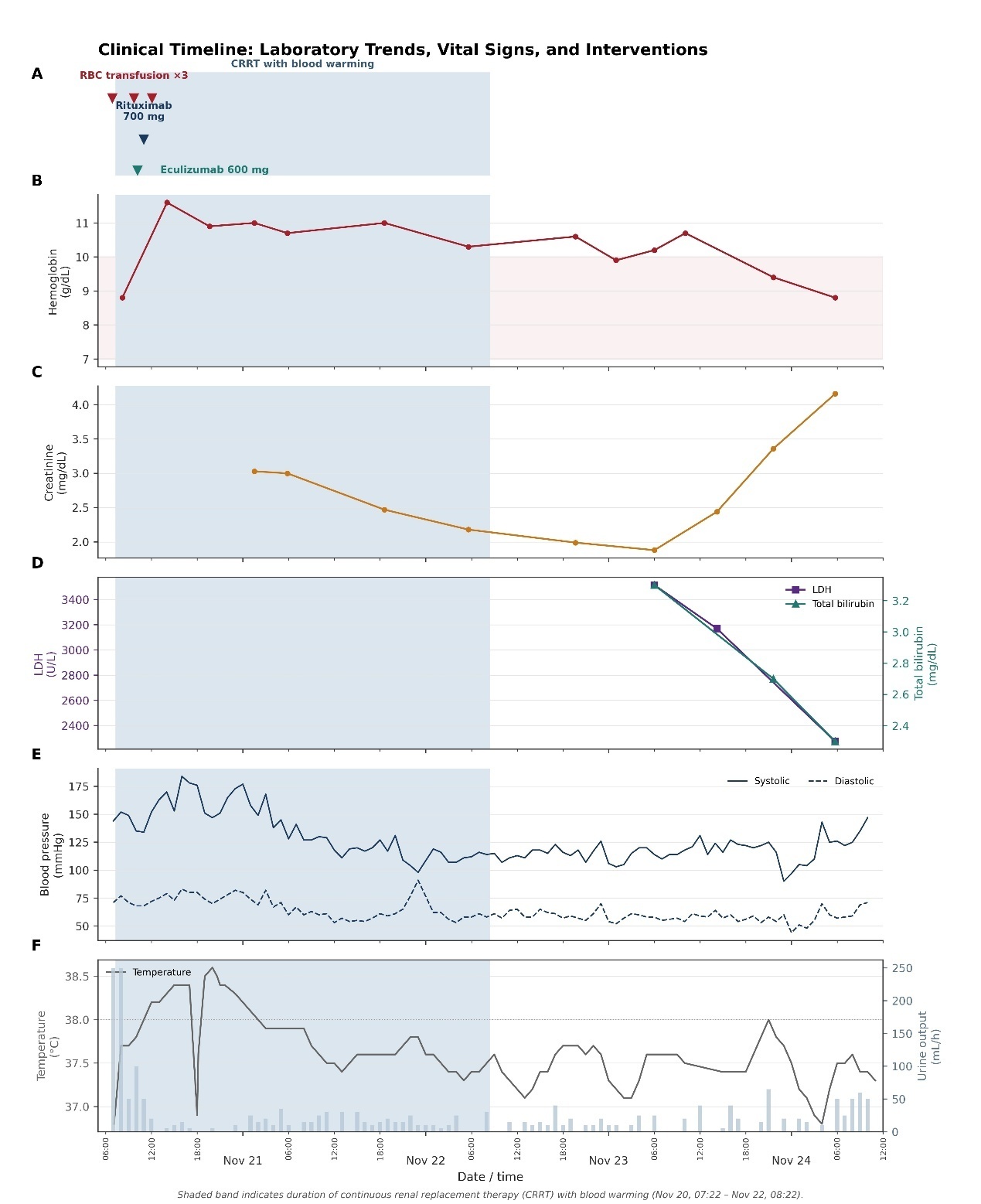
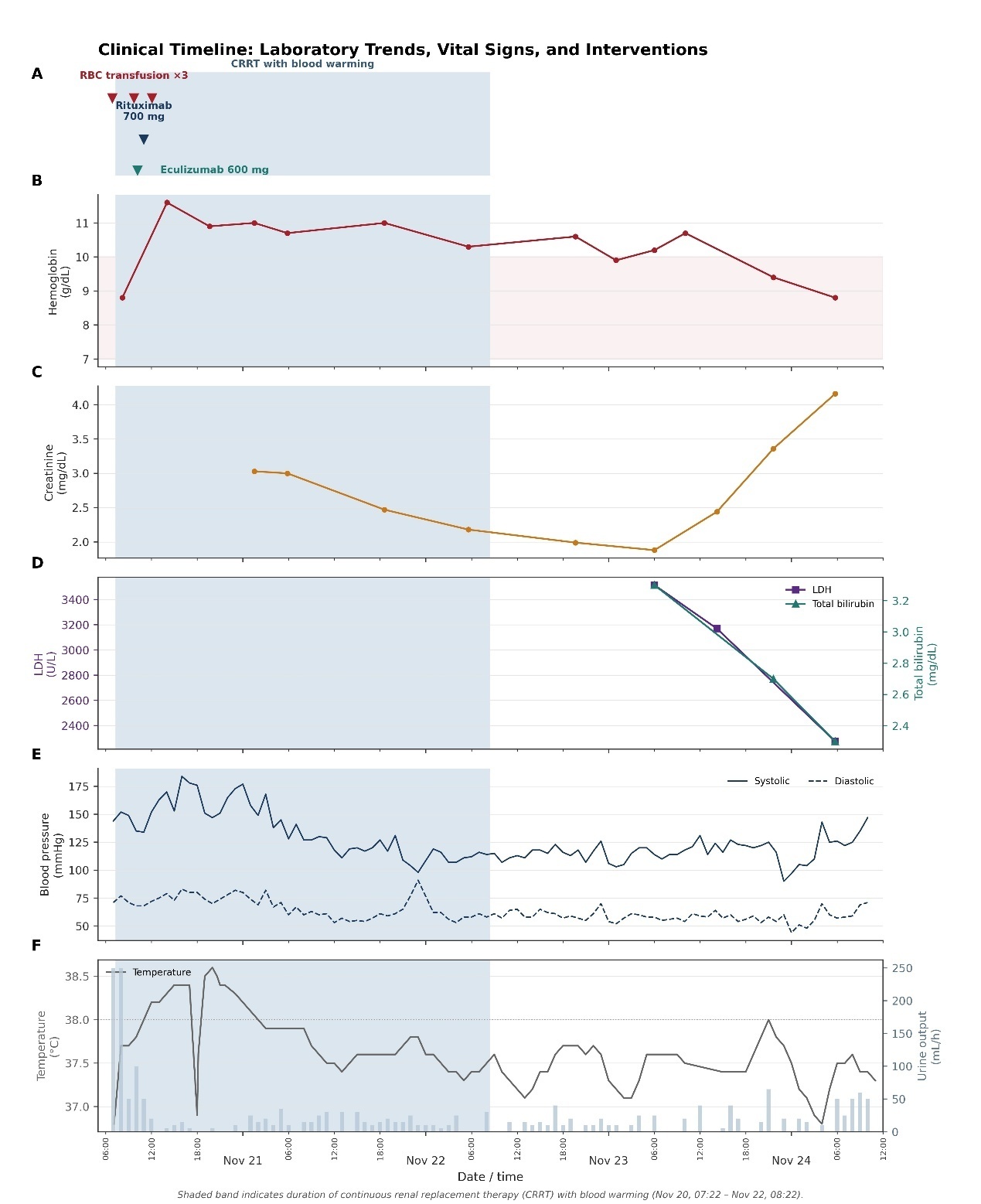
A 3-lumen dialysis central venous catheter, 16 cm, 12 fr, was inserted in the patient’s right jugular vein. The patient was connected to Prismaflex® two hours following ICU admission. Initially the Prismaflex® was set with dialysate rate and replacement rate of zero, with blood flow of 100 ml/min (with the purpose of blood warming alone). The patient was connected to Prismaflex® for 72 hours. At the first 36 hours, Prismaflex® was set with dialysate and replacement rate of zero, but during the following 36 hours it was set at continuous veno-veno hemodialofiltration (CVVHDF) mode, at an effluent dose of 25 ml/kg/hour, with dialysate and replacement rate of 750 ml/hour each and fluid removal rate of 100 ml/hour, due to increasing blood urea level (above 100 mg/dL), creatinine level of 3 mg/dL and oliguria with suspected patient volume overload. Therefore, the total duration of therapy was 72 hours, with Prismaflex® therapy initiated during the first 36 hours for blood warming only.
Citrate was used as an anticoagulant. We set the Prismaflex® temperature to 41ºC and achieved a patient core temperature of approximately 38ºC, measured via a urinary catheter in the urinary bladder. Peripheral temperature was monitored via an oral thermometer. The warming device used was Thermax® (made by Baxter, Prismax systems for critical care). Safety alarms were set on the monitor for temperatures over 39 ºC. A temperature of 41 ºC was selected to enable rapid warming, which is within the manufacturer’s operating specifications. Shortly after instituting these measures, the patient’s hemodynamic and respiratory status began to improve. Within a day, laboratory assays could be performed, and results for electrolytes, creatinine, and bilirubin became available. The hemoglobin stabilized, and no further blood transfusions were required. In addition, Rituximab 375 mg/m2 was administered 50 minutes after Eculizumab administration (Figure 1).
An extensive workup to determine the cause of her cold AIHA was unrevealing: EBV, CMV, HIV, HBV, HCV, and Mycoplasma serology and blood cultures were negative; serum immunoglobulins, free light chains, and protein electrophoresis were normal; and a bone marrow biopsy, including lymphocyte immunophenotyping, was normal. The Donath-Landsteiner test was not performed.
C3 and C4 levels were taken after eculizumab administration and were within normal range, reticulocyte count was high (513.8 K/microL), antibodies screen was positive: DCT Gel Final calc was positive, anti IgG negative and anti-C3 positive (+2). When first measurable, LDH level was 3516 U/L and total bilirubin 3.3 mg/dL (indirect 2.4 mg/dL). Peripheral blood smear showed spherocytosis, reticulocytosis and poikilocytosis. While a formal cold agglutinin test was not performed, the patient’s indirect antiglobulin test (IAT) was strongly positive at 4°C and 22°C and negative at 37°C; the precise thermal amplitude was not established because intermediate temperatures were not tested. This might confirm the presence of a cold-reactive antibody in the patient’s plasma with a thermal amplitude of at least 4-22 °C.
Differential diagnosis included acute/transient complement-mediated cold AIHA of undetermined etiology; paroxysmal cold hemoglobinuria, an autoimmune characterized by -mediated intravascular hemolysis after cold exposure; cold agglutinin disease; secondary cold agglutinin syndrome; and drug-induced immune hemolytic anemia. Drug-induced immune hemolytic anemia was considered a possibility. During the days before hospital admission, the patient received penicillin and NSAIDs. However, specialized testing to confirm this diagnosis was not performed.
She required hemodialysis for a number of weeks, and her kidney function gradually recovered, enabling the discontinuation of renal replacement therapy. Her hemoglobin remained normal with no evidence of hemolysis, and her cold agglutinin-mediated test was negative one month after discharge. One year after the acute hemolytic episode, she remains healthy.
Discussion
Management of severe or symptomatic anemia due to cold AIHA has traditionally been based upon red blood cell transfusion and warming the patient. Plasmapheresis may be initiated in refractory cases. Administration of IVIG is controversial.14 Rituximab is usually administered concurrently to suppress autoantibody production; however, it is not expected to immediately suppress hemolysis. Recently, monoclonal antibodies capable of binding activated complement fragments have been developed. Sutimlimab specifically binds and inhibits C1s, the serine protease subcomponent of C1, and is administered intravenously on an ongoing basis in patients with cold agglutinin disease, and may also be effective in abating acute hemolysis. Eculizumab, a C5 inhibitor used in paroxysmal nocturnal hemoglobinuria, a disease characterized by complement-mediated red cell destruction, also has the potential to dampen acute intravascular hemolysis resulting from complement activation. Eculizumab has prospective evidence for reducing hemolysis in cold agglutinin disease but does not prevent upstream C3 opsonization (15), whereas Sutimlimab blocks the classical pathway at C1 (9,12). Since Sutimlimab was not available at our medical center and immediate rescue therapy was required, Eculizumab was administered.
Our patient presented with massive intravascular hemolysis on the basis of a cold-reactive antibody, possibly due to drug exposure following an extensive dental procedure. She developed oliguric renal failure, metabolic acidosis, pulmonary edema/volume overload following repeated transfusions in the setting of acute kidney injury and hemodynamic instability over a number of hours. As an emergency measure to reduce cold-antibody binding, we initiated extracorporeal blood warming using Prismaflex®, a continuous renal replacement therapy device. This resulted in rapid warming of the patient’s circulation, evidenced by core and peripheral body temperature of 38ºC. Three hours later, eculizumab was administered, and Rituximab was administered 50 minutes later. This combination treatment may have contributed to rapid control of hemolysis, as was evidenced by the resolution of acrocyanosis and improvement in hemodynamic parameters shortly after (Figure 1). Blood chemistry parameters that could not previously be analyzed could now be measured successfully. The hemoglobin stabilized without the need for further blood transfusions. Since both interventions were instituted essentially simultaneously, their individual contribution cannot be disentangled. As mentioned, in addition, the patient was administered Rituximab, while withdrawal of the potentially offending drugs, warmed transfusion, generalized warming, and supportive ICU treatment were occurring concurrently.
Another option that was considered as a temporizing measure in this case was plasmapheresis to physically reduce the load of intravascular IgM cold autoantibodies. However, we considered continuous renal replacement therapy without dialysis or filtration preferable because of its immediate availability and lower rate of adverse reactions. Another option for extracorporeal blood warming is extracorporeal membrane oxygenation therapy (ECMO), but it requires the insertion of 2 large-bore intravenous cannulas with full anticoagulation, a much more aggressive, risk-associated, and expensive measure.
In summary, to our knowledge, this is the first report of combined extracorporeal blood warming and complement inhibition as a rescue measure to treat massive cold AIHA. We suggest that this strategy may represent a rescue option in selected life-threatening cases when routine patient warming is insufficient to abrogate the acute hemolytic process, and until definitive therapy becomes effective and the autoimmune process resolves.
Ethical approval
IRB review was not required for a single de-identified case at our institution.
Consent for publication
Patient consent was obtained for the case report.
Data availability statement
The datasets generated during and/or analyzed during the current study are available from the corresponding author upon reasonable request.
Author contributions
Conceptualization: Sara Dichtwald (Lead). Data curation: Sara Dichtwald (Lead). Project administration: Sara Dichtwald (Equal), Avraham Meyer (Equal), Ilana Hellmann (Equal), Nisim Ifrach (Equal), Martin Ellis (Equal). Visualization: Sara Dichtwald (Lead). Writing – original draft: Sara Dichtwald (Lead). Writing – review & editing: Sara Dichtwald (Equal), Avraham Meyer (Equal), Ilana Hellmann (Equal), Nisim Ifrach (Equal), Martin Ellis (Equal).
Funding
none.
Conflict of interests
none.
Acknowledgement
None.
Declaration of generative AI use
AI tools were not used in the preparation of this manuscript.